Category Archives: inflammation

Anti-inflammatory, vegetable chicken soup, nutritional content, short chain fatty acid production, and health benefits

I recently enjoyed an on-line conversation with physicians and scientists about the benefits of an evolutionary biology approach to human nutrition. During the paleolithic period, fiber consumption was estimated to be 100 grams or more per day. This represents 7 times what most Americans consume and 3-4 times the present daily dietary recommendations. I previously posted a recipe for an anti-inflammatory vegetable soup cooked in broth. I have modified that recipe to a slight degree and did an AI search using Gemini to create tables of nutritional content, the resulting expected production of SCFAs (short chain fatty acids) and the resulting health benefits of this recipe. Here are the results.

You may be surprised by the many health benefits of this nutritional content. Through multiple mechanisms consuming this kind of soup will improve insulin sensitivity, glucose and cholesterol control, increase your production of GLP-1 and other incretins, improve sleep and reduce stress hormones, improve gut barrier function, gut health, and calm your brain. All that from some vegetable soup, easy to make, store, and consume every day.

Conversation with Gemini

Assume this recipe is cooked and separated into 9 equal portions, eating one portion per day. Provide a table with the following nutritional content for each of the components and total each at the bottom of the table. Include fiber, both soluble and insoluble, protein, starch, sugar, fat, micronutrients including all vitamins and minerals, polyphenol total and other important micronutrients. Provide the data for a portion as defined by 1/9 of the total recipe. Finally calculate the production of SCFAs from a portion of this recipe. 

3 quarts of organic chicken broth

fresh garlic, 1 head, crush/press/chop/or use food processor to

release important enzymes before cooking (let prepared garlic sit for 10

minutes before cooking, this allows the enzymes released to produce

important micro nutrients, cooking destroys the enzymes) I use a volume of

fresh garlic equal to the fresh ginger

one large piece (4) inches long)

of fresh ginger root, chop or slice then put into food processor

one large red onion, cut into

small pieces

one bunch of green onions, slice

1 bunch of Italian parsley, cut

and use stems, save greens for end of cooking

two large purple yams slice into

half inch chunks4 fresh large beets with greens,

slice beets, use the greens, cut stems into pieces

one head kale, chop stems into

1–2-inch pieces, cut greens into eat-size pieces

one head Swiss chard, chop stems

into 1–2-inch pieces, cut greens into eat-size pieces

1 head red cabbage, sliced

1 head of napa cabbage

3 carrots, cut each carrot into

thirds or fourths

3 stalks of celery chop into 2

inch pieces

1 whole leek, cut the greens into

2-inch pieces (scissors) and slice the white part into 1/2-inch slices

16 ounces of sliced button mushrooms

2 tablespoons turmeric

4 bay leaves, 4 tsp salt

Here is the nutritional breakdown per portion (1/9th of the total cooked recipe) separated into distinct tables.

Table 1: Fiber Profile (per portion)

Ingredient / ComponentSoluble Fiber (g)Insoluble Fiber (g)Total Fiber (g)
Chicken Broth0.00.00.0
Fresh Garlic0.10.10.2
Fresh Ginger0.10.10.2
Red Onion0.10.20.3
Green Onions0.00.10.1
Italian Parsley0.10.10.2
Purple Yams0.30.71.0
Beets + Greens0.30.60.9
Kale0.10.20.3
Swiss Chard0.10.20.3
Red Cabbage0.71.42.1
Napa Cabbage0.40.71.1
Carrots0.20.40.6
Celery0.10.10.2
Leek0.10.10.2
Button Mushrooms0.10.50.6
Turmeric0.10.20.3
Bay Leaves & Salt0.00.10.1
TOTAL2.8 g5.7 g8.5 g

Table 2: Macronutrients & Carbohydrates (per portion)

Ingredient / ComponentProtein (g)Starch (g)Sugar (g)Fat (g)
Chicken Broth2.10.00.40.8
Fresh Garlic0.30.00.10.0
Fresh Ginger0.10.00.10.0
Red Onion0.20.00.70.0
Green Onions0.00.00.00.0
Italian Parsley0.20.00.10.0
Purple Yams0.54.80.20.0
Beets + Greens0.80.01.80.1
Kale0.30.00.20.1
Swiss Chard0.40.00.20.0
Red Cabbage1.40.03.80.2
Napa Cabbage1.10.01.30.2
Carrots0.20.00.90.0
Celery0.10.00.20.0
Leek0.10.00.30.0
Button Mushrooms1.60.01.00.2
Turmeric0.10.30.00.1
Bay Leaves & Salt0.00.00.00.0
TOTAL9.5 g5.1 g11.1 g1.7 g

Table 3: Vitamin Content Key Highlights (per portion)

Ingredient / ComponentDominant Vitamins Provided
Chicken BrothNiacin (B3), Vitamin B6
Fresh GarlicVitamin C, Vitamin B6
Fresh GingerVitamin B6, Niacin (B3)
Red OnionVitamin C, Folate (B9)
Green OnionsVitamin K, Vitamin A
Italian ParsleyVitamin K, Vitamin C, Vitamin A, Folate (B9)
Purple YamsVitamin C, Vitamin B6
Beets + GreensFolate (B9), Vitamin A, Vitamin C
KaleVitamin K, Vitamin C, Vitamin A
Swiss ChardVitamin K, Vitamin A, Vitamin C
Red CabbageVitamin C, Vitamin K, Vitamin B6
Napa CabbageVitamin C, Folate (B9), Vitamin K
CarrotsVitamin A (Beta-carotene), Vitamin K, Vitamin B6
CeleryVitamin K, Folate (B9)
LeekVitamin K, Vitamin C, Folate (B9)
Button MushroomsRiboflavin (B2), Niacin (B3), Pantothenic Acid (B5)
TurmericTraces of Vitamin C, Vitamin B6
Bay Leaves & SaltVitamin A, Vitamin C (trace amounts from bay leaves)
TOTAL SUMMARYExtremely rich in Vitamins K, C, A, and B-complex (Folate, B6, Niacin, B2)

Table 4: Mineral Content Key Highlights (per portion)

Ingredient / ComponentKey Minerals Provided
Chicken BrothSodium, Potassium, Phosphorus
Fresh GarlicManganese, Selenium, Calcium
Fresh GingerMagnesium, Potassium, Manganese
Red OnionPotassium, Calcium
Green OnionsCalcium, Potassium
Italian ParsleyIron, Potassium, Calcium
Purple YamsPotassium, Manganese, Copper
Beets + GreensManganese, Potassium, Iron, Magnesium
KaleCalcium, Potassium, Copper
Swiss ChardMagnesium, Potassium, Iron
Red CabbagePotassium, Manganese, Calcium
Napa CabbageCalcium, Potassium
CarrotsPotassium, Manganese
CeleryPotassium, Sodium
LeekManganese, Iron
Button MushroomsSelenium, Copper, Potassium, Phosphorus
TurmericIron, Manganese
Bay Leaves & SaltSodium (~900mg per portion from salt), Magnesium
TOTAL SUMMARYHigh in Potassium, Manganese, Sodium, Iron, Selenium, Calcium, & Magnesium

Calculation of SCFA (Short-Chain Fatty Acid) Production

Short-chain fatty acids (primarily Acetate, Propionate, and Butyrate) are synthesized via colonic microbial fermentation of non-digestible carbohydrates (fermentable soluble fibers, insoluble structural fibers, and resistant starches).

Isabel Smith Nutrition

1. Fermentable Substrate Availability (per portion):

  • Soluble Fiber: ~2.8 g (Nearly 100% fermentable)
  • Insoluble Fiber: ~5.7 g (~20–30% fermentable in brassicas/root veggies) → ~ 1.4 g
  • Resistant Starch (RS): If eaten fresh after cooking, RS is low (~0.3 g from yams). If the soup is cooled overnight in the fridge and reheated, retrograde starch formation (RS3) increases this yield to ~1.5 g.
  • Total Fermentable Substrates: ≈2.8 g+1.4 g+0.3 g to 1.5 g=4.5 g to 5.7 g

2. Yield Assumptions: Human metabolic and gut microbiome studies establish that 1 gram of fermentable substrate yields approximately 8 to 10 mmol of total SCFAs (or roughly 0.4 to 0.5 grams of short-chain fatty acids).

3. Estimated Total SCFA Yield:

SCFA (mmol)=(4.5 to 5.7 grams)×8.5 mmol/g≈38 to 48 mmol per portion

SCFA (mass)≈2.2 to 2.8 grams of total SCFAs per portion

4. Typical SCFA Ratio Breakdown:

  • Acetate (~60%): ≈23 to 29 mmol (Energy substrate for peripheral tissues)
  • Propionate (~20%): ≈7.5 to 9.5 mmol (Utilized in hepatic gluconeogenesis)
  • Butyrate (~20%): ≈7.5 to 9.5 mmol (Primary fuel source for colonocytes and gut barrier repair)

(Tip: Letting the cooked soup cool in the refrigerator overnight before portioning maximizes resistant starch content, maximizing the butyrate yield).

Assume the soup is cooled overnight and then reheated, what is the SCFA production?

Cooling the cooked soup overnight triggers starch retrogradation, converting digestible starches (specifically the amylose chains from the purple yams) into Type-3 Resistant Starch (RS3). Reheating the soup at normal eating temperatures retains the vast majority of this crystalline RS3 structure.

GlobalRPH+ 1

Because RS3 resists digestion in the small intestine, it travels into the large intestine where colonic bacteria ferment it into additional Short-Chain Fatty Acids (SCFAs).

GlobalRPH

1. Shift in Fermentable Substrates per Portion

  • Soluble Fiber: ~2.8 g (~100% fermentable)
  • Fermentable Insoluble Fiber: ~1.4 g (~25% of total insoluble fiber)
  • Resistant Starch (RS3): ~1.8 to 2.4 g (up from ~0.3 g when freshly cooked, as ~35–50% of the yam starch retrogrades)
  • Total Fermentable Substrates: ~6.0 to 6.6 grams per portion

2. Recalculated SCFA Production per Portion

Based on standard colonic fermentation yields (~8.5 mmol of total SCFA produced per gram of fermented substrate):

Total SCFA (mmol)=(6.0 to 6.6 g)×8.5 mmol/g≈51 to 56 mmol per portion

Total SCFA (mass)≈3.0 to 3.3 grams of total SCFAs per portion

This represents an approximate 20–35% increase in total SCFA output compared to eating the soup immediately after cooking.

3. SCFA Breakdown by Type

Resistant starch (RS3) specifically favors butyrate-producing bacteria (such as Faecalibacterium prausnitzii and Eubacterium rectale), slightly shifting the overall SCFA balance:

  • Acetate (~55%): ≈28 to 31 mmol
    • Function: Enters systemic circulation; fuels muscle, heart, and brain tissue.
  • Propionate (~20%): ≈10 to 11 mmol
    • Function: Travels to the liver; involved in gluconeogenesis and satiety signaling.

GlobalRPH

  • Butyrate (~25%): ≈13 to 14 mmol
    • Function: Primary energy source for colonocytes (gut lining cells), reduces inflammation, and strengthens the mucosal barrier.

Blue Horizon Blood Tests

Describe the health benefits of these SCFAs, including but not limited to glucose control, cholesterol, appetite suppression, satiety, insulin sensitivity, anti-inflammatory properties, gut barrier function, colonic health, regularity, sleep and stress.

Short-chain fatty acids (SCFAs)—primarily acetate, propionate, and butyrate—act as vital signaling molecules throughout your body, linking gut microbial activity directly to systemic metabolic, immune, and neurological health.

1. Gut Barrier Function & Colonic Health

  • Fuel for Colonocytes: Butyrate serves as the primary energy source for the epithelial cells lining your colon, supplying up to 70% of their total ATP requirements.
  • Tight Junction Integrity: Butyrate and propionate upregulate the expression of key tight-junction proteins (claudin-1, occludin, and ZO-1). This seals the intestinal epithelial layer, preventing “leaky gut” and stopping bacterial lipopolysaccharides (LPS) from entering circulation.
  • Mucus Layer Maintenance: SCFAs stimulate goblet cells to produce MUC2, reinforcing the protective gel-like mucus layer that shields gut tissue from physical and chemical damage.

2. Regularity & Digestive Motility

  • Smooth Muscle Stimulation: SCFAs interact with free fatty acid receptors (FFAR2 and FFAR3) on enteric neurons to stimulate peristaltic reflexes, accelerating colonic transit time and encouraging regular bowel movements.
  • Osmotic Balance: Microbial fermentation alters local pH and osmotic pressure, attracting sufficient water to the stool to prevent constipation without causing osmotic diarrhea.

3. Glucose Control & Insulin Sensitivity

  • Incretin Hormone Release: Propionate and butyrate trigger L-cells in the distal intestine to secrete GLP-1 (Glucagon-Like Peptide-1) and PYY (Peptide YY). GLP-1 enhances glucose-dependent insulin secretion from the pancreas while suppressing glucagon.
  • Skeletal Muscle Glucose Uptake: Acetate and butyrate activate AMPK (AMP-activated protein kinase) in skeletal muscle, increasing GLUT4 transporter translocation to muscle cell membranes, which boosts insulin-independent glucose clearance.
  • Hepatic Gluconeogenesis Regulation: Propionate travels to the liver via the portal vein where it regulates hepatic glucose production, preventing excessive overnight glycogen breakdown.

4. Cholesterol & Lipid Metabolism

  • Inhibition of Cholesterol Synthesis: Propionate inhibits HMG-CoA reductase, the rate-limiting enzyme in liver cholesterol production, helping lower circulating total and LDL cholesterol levels.
  • Upregulation of Bile Acid Excretion: SCFAs lower colonic pH, promoting the conversion of primary bile acids into secondary bile acids, which binds dietary cholesterol and increases its excretion through stool.

5. Appetite Suppression & Satiety

  • Central Brain Signaling: GLP-1 and PYY secreted in response to SCFAs cross the blood-brain barrier to target the hypothalamus, signaling satiety and reducing food intake.
  • Direct Acetate Signaling: Acetate crosses the blood-brain barrier directly and accumulates in the hypothalamus, altering the expression of neuropeptides (POMC/CART) that suppress appetite and reduce cravings.

6. Anti-Inflammatory & Immune Properties

  • HDAC Inhibition: Butyrate acts as a natural Histone Deacetylase (HDAC) inhibitor. By inhibiting HDACs, it upregulates anti-inflammatory gene expression and suppresses pro-inflammatory cytokines like TNF-alpha, IL-6, and IL-1β.
  • T-Regulatory Cell Induction: SCFAs promote the differentiation of naive T-cells into Treg (regulatory T-cells) in the gut-associated lymphoid tissue (GALT), helping prevent autoimmune reactions, food allergies, and inflammatory bowel disease (IBD).

7. Stress, Sleep, & The Gut-Brain Axis

  • Vagus Nerve Activation: SCFAs bind to receptors on the vagus nerve terminal in the gut wall, sending direct inhibitory signals to the central nervous system to dampen stress reactivity.
  • HPA Axis Modulation: By preserving gut barrier integrity and preventing low-grade systemic inflammation (endotoxemia), SCFAs lower baseline cortisol levels and reduce hypothalamic-pituitary-adrenal (HPA) axis overdrive.
  • Neurotransmitter Synthesis & Sleep Architecture: Butyrate and propionate encourage enterochromaffin cells to produce serotonin (the precursor to melatonin) and GABA. Higher SCFA levels promote deeper, more stable slow-wave sleep (NREM stage 3) by calming central nervous system excitability.

How does the SCFA yield and prebiotic diversity of this soup compare to taking a daily commercial prebiotic supplement?

A single daily portion of this cooked-and-cooled vegetable soup outperforms most single-ingredient commercial prebiotic supplements in both total yield and structural diversity.

Direct Comparison: Soup Portion vs. Typical Commercial Prebiotic

AttributeSoup Portion (1/9th Recipe, Cooled & Reheated)Standard Commercial Prebiotic (e.g., Inulin/GOS Powder)
Total Prebiotic Yield~6.0 to 6.6 g per portion (Soluble fiber, RS3, oligosaccharides)~3.0 to 5.0 g per serving (Standard scoop/capsule dose)
SCFA Production~51 to 56 mmol (~3.0 to 3.3 g total SCFAs)~25 to 45 mmol (Varies based on single-substrate fermentation rate)
Substrate Diversity7+ Distinct Prebiotic Types1 to 2 Substrates (Usually single isolated fiber type)
Microbiome SpreadFerments across Upper, Mid, and Lower ColonFerments rapidly in Proximal (Upper) Colon only
TolerabilityHigh (Diverse fibers ferment slowly over time)Moderate to Low (Single isolated fibers often cause rapid gas/bloating)

Key Advantages of Whole-Food Soup Diversity

1. Substrate Complexity Prevents Gut Monoculture

Commercial prebiotics typically rely on isolated fibers like inulin, fructooligosaccharides (FOS), or galactooligosaccharides (GOS). While effective, a single substrate selectively feeds only 1–2 specific bacterial species (such as Bifidobacteria).

The soup provides a complex mixture of distinct fibers that nourish a broader, healthier ecosystem:

  • Inulin & FOS (Garlic, onions, leeks) → Bifidobacterium
  • Beta-Glucans (Button mushrooms) → Lactobacillus and macrophage-activating immune cells
  • Type-3 Resistant Starch / RS3 (Cooled purple yams) → Faecalibacterium prausnitzii (primary butyrate producer)
  • Pectin & Mucilages (Carrots, beets) → Akkermansia muciniphila and Bacteroides
  • Arabinogalactans & Cellulose (Brassicas: kale, cabbages, Swiss chard) → Roseburia and Ruminococcus

2. Full-Length Colonic Fermentation

Isolated prebiotic supplements (especially inulin) ferment very rapidly in the proximal (beginning) colon. This rapid gas production often leads to uncomfortable bloating, while leaving the distal (end) colon under-nourished.

Because the soup combines fast-fermenting soluble fibers with slow-fermenting insoluble structures and resistant starches, fermentation occurs gradually along the entire length of the large intestine. This ensures distal colonocytes receive a steady supply of butyrate, which is critical because the distal colon is where most colonic pathologies (like ulcerative colitis and colorectal cancers) typically develop.

3. Synergistic Bioactive Matrix

Commercial supplements supply isolated fiber without the supporting food matrix. The soup delivers prebiotics alongside polyphenols (curcumin, quercetin, anthocyanins) and glucosinolates.

Microbial enzymes break down these complex polyphenols into secondary active metabolites, while the polyphenols simultaneously act as “selective antimicrobials,” inhibiting pathogenic bacteria (Clostridia, E. coli) so beneficial SCFA producers can thrive.

Eat clean, live clean, sleep well, exercise wisely, rest often, enjoy the company of loved ones, spend time outdoors and live in the present.

Bob Hansen MD
What can I do to improve insulin sensitivity?

  1. Exercise (aerobic and resistance training, in the gym, on the dance floor, outside in a greenspace, with friends and family)
  2. Get adequate restorative sleep (follow regular sleep habits, see also #s1, 3, 4 to 12)
  3. Enhance circadian rhythm (outdoor light exposure early in the day, avoid screen time prior to bed and/or use blue light blocking glasses and screen software, see also #1, #2, # 4 to 12)
  4. Reduce stress (meditation, yoga, tai chi, family time, #s1-3, 5-12)
  5. Engage in meaningful work
  6. Spend time with those you love
  7. Eat an anti-inflammatory (paleo) diet
  8. If overweight, employ therapeutic carbohydrate restriction (if on medications for diabetes this must be done under medical supervision following established published guidelines for medication adjustments)
  9. Spend time outdoors in a greenspace, especially early in the day. (Forest bathing)
  10. When working (on the computer) or reading indoors, do it in front of a large window to increase outdoor light exposure.
  11. Regularly use a sauna
  12. Consider brief cold immersion sessions (2-4 minutes of cold shower or cold-water immersion, approach this gradually)

Exercise: per minute spent, resistance training offers the most benefit. Second to resistance training is HIIT (High Intensity Interval Training) per unit of time spent. Finally moderate aerobic exercise (heart rate 60 to 80% of maximum predicted heart rate for age) places third for benefit per unit time spent. Most importantly, find some exercise that you enjoy and will sustain. Dancing, especially for seniors, combines the benefits of socialization, exercise, and simultaneous use of multiple brain areas, preserving and enhancing cognitive function in addition to mitigation of insulin resistance, chronic inflammation and cardiovascular risk. Engaging in sports that require coordination, balance, strength and complex movements also utilizes multiple areas of the brain simultaneously, providing for cognitive, metabolic and cardiovascular benefit.  A single bout of exercise can increase insulin sensitivity for at least 16 h post exercise in healthy as well as diabetic subjects.

Sleep and Circadian Rhythm

Just one night of short-sleep causes acute insulin resistance. Chronic sleep deprivation contributes to chronic inflammation and insulin resistance. Go the bed and wake up on a consistent schedule. Avoid food and screen time for 3 hours before bed. Allow yourself at least 8 hours per night of sleep opportunity. Find relaxing routines to follow for an hour or two before bedtime. Avoid strenuous exercise in the evening. No caffeine after 12 noon. Getting outdoor light exposure early in the day facilitates restorative sleep. During sleep our brains flush out metabolic debris that accumulates during the day through the glymphatic system.  Melatonin production during sleep provides multiple benefits including anti-inflammatory effects. Sleep in a cool very dark bedroom.

Optimizing sleep and circadian rhythm involves a combination of timing, environmental control, and biological signaling. These habits aim to align your internal master clock (the suprachiasmatic nucleus) with the external 24-hour light-dark cycle.

Summary Table: Daily Habits

HabitTimingPrimary Benefit
View SunlightFirst 60 mins of daySets the circadian “timer”
ExerciseMorning or AfternoonIncreases sleep pressure/adenosine
Last Meal3 hours before bedPrevents metabolic circadian shift
Cool RoomAll nightFacilitates core temp drop
Darkness1–2 hours before bedAllows natural melatonin rise

In my next posts I will explore items 5 through 12 above.

Yan R, Chen L, Lin G, Shi Y, Huang W, Mai Y, Sun J, Li D. Comparative effectiveness of different exercise modality on glycaemic control and lipid profile for prediabetes: systematic review and network meta-analysis. Front Endocrinol (Lausanne). 2025 Jul 24;16:1518871. doi: 10.3389/fendo.2025.1518871. PMID: 40778279; PMCID: PMC12328182. https://pubmed.ncbi.nlm.nih.gov/40778279/

Zhang L, Cheng X, Yang Y, Li X, Yuan Y. Optimal dosage and modality of exercise on glycemic control in people with prediabetes: a systematic review and network meta-analysis. Front Endocrinol (Lausanne). 2025 Apr 28;16:1560676. doi: 10.3389/fendo.2025.1560676. PMID: 40357204; PMCID: PMC12066256. https://pubmed.ncbi.nlm.nih.gov/40357204/

Huang L, Fang Y, Tang L. Comparisons of different exercise interventions on glycemic control and insulin resistance in prediabetes: a network meta-analysis. BMC Endocr Disord. 2021 Sep 6;21(1):181. doi: 10.1186/s12902-021-00846-y. PMID: 34488728; PMCID: PMC8422751. https://pubmed.ncbi.nlm.nih.gov/34488728/

Amaravadi SK, Maiya GA, K V, Shastry BA. Effectiveness of structured exercise program on insulin resistance and quality of life in type 2 diabetes mellitus-A randomized controlled trial. PLoS One. 2024 May 21;19(5):e0302831. doi: 10.1371/journal.pone.0302831. PMID: 38771888; PMCID: PMC11108169. https://pubmed.ncbi.nlm.nih.gov/38771888/

Alghadir AH, Gabr SA, Iqbal A. The effects of supervised aerobic training on dyslipidaemia among diabetic older patients. BMC Endocr Disord. 2024 Oct 9;24(1):212. doi: 10.1186/s12902-024-01745-8. PMID: 39385223; PMCID: PMC11462724. https://pubmed.ncbi.nlm.nih.gov/39385223/

Abdelbasset WK. Resistance Exercise Versus Aerobic Exercise Combined with Metformin Therapy in the Treatment of type 2 Diabetes: A 12-Week Comparative Clinical Study. Endocr Metab Immune Disord Drug Targets. 2021;21(8):1531-1536. doi: 10.2174/1871530320999200918143227. PMID: 32957900. https://pubmed.ncbi.nlm.nih.gov/32957900/

Li J, Cheng W, Ma H. A Comparative Study of Health Efficacy Indicators in Subjects with T2DM Applying Power Cycling to 12 Weeks of Low-Volume High-Intensity Interval Training and Moderate-Intensity Continuous Training. J Diabetes Res. 2022 Jan 13;2022:9273830. doi: 10.1155/2022/9273830. PMID: 35071605; PMCID: PMC8776485. https://pubmed.ncbi.nlm.nih.gov/35071605/

Findikoglu G, Altinkapak A, Yaylali GF. Is isoenergetic high-intensity interval exercise superior to moderate-intensity continuous exercise for cardiometabolic risk factors in individuals with type 2 diabetes mellitus? A single-blinded randomized controlled study. Eur J Sport Sci. 2023 Oct;23(10):2086-2097. doi: https://pubmed.ncbi.nlm.nih.gov/36622777/

Cox ER, Gajanand T, Keating SE, Hordern MD, Burton NW, Green DJ, Ramos JS, Ramos MV, Fassett RG, Cox SV, Coombes JS, Bailey TG. Effect of low-volume combined aerobic and resistance high-intensity interval training on vascular health in people with type 2 diabetes: a randomised controlled trial. Eur J Appl Physiol. 2024 Sep;124(9):2819-2833. doi: 10.1007/s00421-024-05473-8. Epub 2024 May 2. PMID: 38695912; PMCID: PMC11365856. https://pubmed.ncbi.nlm.nih.gov/38695912/

Li Z, Luo S, Bai X, Huang L, Guo H, Chen S, Wang D. Effects of different exercise types on vascular endothelial function in individuals with abnormal glycaemic control: a systematic review and network meta-analysis. PeerJ. 2025 Aug 8;13:e19839. doi: 10.7717/peerj.19839. PMID: 40792010; PMCID: PMC12338059. https://pubmed.ncbi.nlm.nih.gov/40792010/

Borghouts LB, Keizer HA. Exercise and insulin sensitivity: a review. Int J Sports Med. 2000 Jan;21(1):1-12. doi: 10.1055/s-2000-8847. PMID: 10683091. https://pubmed.ncbi.nlm.nih.gov/10683091/

Wojtaszewski JF, Richter EA. Effects of acute exercise and training on insulin action and sensitivity: focus on molecular mechanisms in muscle. Essays Biochem. 2006;42:31-46. doi: 10.1042/bse0420031. PMID: 17144878. https://pubmed.ncbi.nlm.nih.gov/17144878/

Böhm A, Weigert C, Staiger H, Häring HU. Exercise and diabetes: relevance and causes for response variability. Endocrine. 2016 Mar;51(3):390-401. doi: 10.1007/s12020-015-0792-6. Epub 2015 Dec 7. PMID: 26643313; PMCID: PMC4762932. https://pubmed.ncbi.nlm.nih.gov/26643313/

Reutrakul S, Van Cauter E. Sleep influences on obesity, insulin resistance, and risk of type 2 diabetes. Metabolism. 2018 Jul;84:56-66. doi: 10.1016/j.metabol.2018.02.010. Epub 2018 Mar 3. PMID: 29510179. https://pubmed.ncbi.nlm.nih.gov/29510179/

Antza C, Kostopoulos G, Mostafa S, Nirantharakumar K, Tahrani A. The links between sleep duration, obesity and type 2 diabetes mellitus. J Endocrinol. 2021 Dec 13;252(2):125-141. doi: 10.1530/JOE-21-0155. PMID: 34779405; PMCID: PMC8679843. https://pubmed.ncbi.nlm.nih.gov/34779405/

Koren D, Taveras EM. Association of sleep disturbances with obesity, insulin resistance and the metabolic syndrome. Metabolism. 2018 Jul;84:67-75. doi: 10.1016/j.metabol.2018.04.001. Epub 2018 Apr 6. PMID: 29630921. https://pubmed.ncbi.nlm.nih.gov/29630921/

Duffy, J. F., & Czeisler, C. A. (2009). Effect of Light on Human Circadian Physiology. Sleep Medicine Clinics. https://pubmed.ncbi.nlm.nih.gov/20161220/

Okamoto-Mizuno, K., & Mizuno, K. (2012). Effects of thermal environment on sleep and circadian rhythm. Journal of Physiological Anthropology. https://pubmed.ncbi.nlm.nih.gov/22738673/

Roenneberg, T., et al. (2012). Social Jetlag and Obesity. Current Biology. https://pubmed.ncbi.nlm.nih.gov/22578422/

Drake, C., et al. (2013). Caffeine effects on sleep taken 0, 3, or 6 hours before going to bed. Journal of Clinical Sleep Medicine. https://pubmed.ncbi.nlm.nih.gov/24235903/

Gooley, J. J., et al. (2011). Exposure to Room Light before Bedtime Suppresses Melatonin Onset and Shortens Melatonin Duration in Humans. The Journal of Clinical Endocrinology & Metabolism. https://pubmed.ncbi.nlm.nih.gov/21193540/

Speksnijder EM, Bisschop PH, Siegelaar SE, Stenvers DJ, Kalsbeek A. Circadian desynchrony and glucose metabolism. J Pineal Res. 2024 May;76(4):e12956. doi: 10.1111/jpi.12956. PMID: 38695262. https://pubmed.ncbi.nlm.nih.gov/38695262/

Engin A. Misalignment of Circadian Rhythms in Diet-Induced Obesity. Adv Exp Med Biol. 2024;1460:27-71. doi: 10.1007/978-3-031-63657-8_2. PMID: 39287848. https://pubmed.ncbi.nlm.nih.gov/39287848/

Koh JYJ, Tan CYH, Li M, Liu MH, Chew HSJ. The Effectiveness of Time-Restricted Eating as an Intermittent Fasting Approach on Shift Workers’ Glucose Metabolism: A Systematic Review and Meta-Analysis. Nutrients. 2025 May 15;17(10):1689. doi: 10.3390/nu17101689. PMID: 40431429; PMCID: PMC12114545. https://pubmed.ncbi.nlm.nih.gov/40431429/

Wang A, Vreijling J, Jongejan A, Rumanova VS, Versteeg RI, Kalsbeek A, Serlie MJ, la Fleur SE, Bisschop PH, Baas F, Stenvers DJ. The Acute Effects of Morning Bright Light on the Human White Adipose Tissue Transcriptome: Exploratory Post Hoc Analysis. Clocks Sleep. 2025 Aug 27;7(3):45. doi: 10.3390/clockssleep7030045. PMID: 40981208; PMCID: PMC12452623. https://pubmed.ncbi.nlm.nih.gov/40981208/

THIS WEBSITE PROVIDES INFORMATION FOR EDUCATIONAL PURPOSES ONLY. CONSULT YOUR HEALTH CARE PROVIDER FOR MEDICAL ADVICE.

Eat clean, drink filtered water, love, laugh, exercise outdoors in a greenspace, get some morning sunlight, block the blue light before bed, engage in meaningful work, find a sense of purpose, spend time with those you love, AND sleep well tonight.

Doctor Bob

Insulin Resistance, the silent killer and root cause of modern chronic disease.

Insulin is much more than a blood sugar hormone. Produced by the pancreas primarily in response to carbohydrate and sugar consumption, insulin is a master anabolic signal that dictates how every cell in your body grows, uses energy, and repairs itself. When insulin levels are healthy, it keeps the body in a state of “build and store.” When insulin resistance (IR) develops, the body loses its ability to hear this signal, leading to systemic breakdown. Instead of “build and store” the body deteriorates, causing loss of muscle mass, strength, energy production, memory and cognitive function, bone strength, brain cells and connections, ability of blood vessels to relax, ability for the heart to pump blood, ability to achieve restorative sleep, ability of the liver and kidneys to clear toxins from the body, even the ability to reproduce resulting in infertility and erectile dysfunction. Visceral fat stores increase to destructive levels resulting in obesity and obesity-related complications including chronic inflammation which further drives IR to higher levels.

IR is a root cause of cardiovascular disease (heart attack, stroke, hypertension, heart failure), many kinds of cancer (directly linked to breast, prostate and colon cancer), kidney failure, heart failure, dementia, osteoporosis, osteoarthritis, and much more.  IR is causally linked or a contributor to, every chronic non-communicable disease of modern civilization.

WHAT IS INSULIN RESISTANCE?

Insulin resistance is the inability of cells and organs to respond normally to insulin signaling. Every cell of every organ has insulin receptors that initiate action by the cell and organ.

WHAT CAUSES INSULIN RESISTANCE?

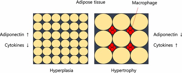
There are many causes of IR. Stress hormones (cortisol, adrenaline), inflammation, and high insulin levels themselves (response to dietary sugar and refined carbohydrates), each alone and in combination, cause immediate (within minutes to hours) insulin resistance. When these conditions persist over time insulin resistance becomes a chronic state. As fat cells grow in size, they reach a point where there is inadequate blood flow to the cells themselves and macrophages (immune cells that reside between the fat cells, most prominently in visceral fat) produce inflammatory chemicals called cytokines. Cytokines flow through the blood stream and effect every organ and every cell in the body creating a state of chronic inflammation which further worsens IR, creating a vicious cycle. As IR continues the pancreas produces increasingly higher amounts of insulin to maintain normal blood sugar levels but eventually IR becomes so great that blood sugar levels move into the “pre-diabetes” and eventually the diabetes range. IR builds for years to decades before blood sugar regulation fails. By the time blood sugar levels are “abnormal” insulin resistance has done great damage throughout the body.

Most doctors tragically do not order fasting insulin levels as routine blood tests. Fasting insulin levels rise long before fasting blood sugars and hemoglobin A1c start to rise. Meanwhile the damage progresses under the radar of routine testing.


1. Metabolic Engines: Muscle and Liver

Muscle

  • Normal Action: Insulin acts as a key that opens “doors” (GLUT4 receptors) to let glucose in for fuel. it also stimulates protein synthesis. Protein synthesis is essential to maintaining and increasing muscle mass and strength.
  • Insulin Resistance Effect: The “doors” stay locked. Glucose stays in the blood, and the muscle becomes “starched,” leading to sarcopenia (muscle wasting) and fatigue. The muscle can no longer utilize dietary protein to maintain or increase muscle mass.

Liver

  • Normal Action: Tells the liver to stop producing glucose and start storing it as glycogen or converting excess into fat.
  • Insulin Resistance Effect: The liver ignores the “stop” signal and keeps pumping out glucose while simultaneously ramping up fat production. This results in Non-Alcoholic Fatty Liver Disease (NAFLD).

2. Fat Cells (Adipose Tissue)

Visceral (Deep Fat) vs. Subcutaneous (Under Skin)

  • Normal Action: Insulin promotes fat storage and inhibits the breakdown of stored fat (lipolysis).
  • Insulin Resistance Effect: Fat cells—especially visceral ones—become “leaky.” They spill free fatty acids into the bloodstream and release inflammatory cytokines. This causes weight gain that is biologically difficult to lose because high insulin levels keep the “fat-burning” switch permanently off.

3. The Vital Organs: Heart, Kidneys, and Arteries

Heart and Arteries

  • Normal Action: Insulin stimulates the release of nitric oxide, which helps arteries relax and dilate.
  • Insulin Resistance Effect: Nitric oxide production drops, causing arteries to stiffen (hypertension). High insulin also damages the endothelial lining, leading to atherosclerosis (plaque buildup). This is the primary driver of heart failure, heart attacks and strokes.

Kidneys

  • Normal Action: Helps regulate sodium reabsorption.
  • Insulin Resistance Effect: The kidneys hold onto too much salt, increasing blood pressure. Over time, high blood sugar and inflammation damage the filtering units, leading to chronic kidney disease (CKD).

4. The Brain, Memory, and Sleep

Brain and Memory

  • Normal Action: Insulin crosses the blood-brain barrier to regulate appetite and support synaptic plasticity (the basis of learning).
  • Insulin Resistance Effect: Often called “Type 3 Diabetes,” brain IR starves neurons of energy and allows amyloid plaques and neurofibrillary tangles to build up. Worse, the brain is unable to utilize glucose to meet energy demands it starts to malfunction. This is a direct pathway to Alzheimer’s disease and dementia. As the small arteries in the brain become atherosclerotic and unable to deliver adequate oxygen and nourishment to brain cells small areas of the brain become permanently damaged eventually leading to vascular dementia.

Sleep

  • Insulin Resistance Effect: IR is heavily linked to Obstructive Sleep Apnea. (OSA) High insulin affects the central respiratory drive and increases fat deposits around the neck (a major contributor to obstructive sleep apnea), disrupting sleep cycles and creating periods of inadequate oxygen flow to the brain resulting in the acute stress response and awakening with each apneic event. Even without OSA, high insulin levels impair the production of melatonin which is essential to normal-restorative sleep. Throughout the day the brain accumulates metabolic toxins that must be cleared through the glymphatic system at night during sleep. As sleep is impaired this clearance system is disrupted, contributing to structural damage and functional loss. Sleep disruption and apneic episodes are stressful events, increasing stress hormones which then worsen IR, creating another vicious cycle. One night of sleep disruption causes acute IR. Chronic sleep disruption contributes to chronic IR.

5. Immunity and Structural Health

Immune System

  • Action: High insulin/glucose impairs white blood cell function.
  • Effect: Chronic inflammation (high CRP levels) and a weakened defense against infections. This is why diabetics often have poor wound healing. As normal immune regulation is impaired the immune system both over-reacts and under-reacts. Under-reaction increases risk of infection. Over-reaction produces cytokine storms seen with Covid-19 and other infections. Chronic inflammation worsens IR creating another vicious cycle. Chronic inflammation contributes to most chronic diseases.

Bone and Joints

  • Action: Insulin is bone-building.
  • Effect: IR leads to poor bone quality (despite high density) and osteoarthritis due to systemic inflammation and the “glycosylation” (sugar-coating) of joint cartilage, making it brittle.

6. The Pancreas: Beta and Alpha Cells

  • Normal Action: Beta cells produce insulin; Alpha cells produce glucagon (which raises sugar). They balance each other.
  • Insulin Resistance Effect:
    • Beta Cells: Work overtime to produce massive amounts of insulin to compensate, eventually “burning out” and dying. This can produce per4manent irreversible damage to the pancreas.
    • Alpha Cells: Become resistant to insulin’s “stop” signal and keep secreting glucagon, further raising blood sugar levels which in turn cause higher insulin secretion, both of which worsen IR, creating another vicious cycle.

7. Reproductive Effects: Infertility

  • In Women: High insulin stimulates the ovaries to produce excess testosterone, which is the primary driver of Polycystic Ovary Syndrome (PCOS) and infertility.
  • In Men: IR is a leading cause of low testosterone and erectile dysfunction (due to the arterial damage mentioned above).

Summary of Systemic Effects

ConditionPrimary Mechanism of Insulin Resistance
AtherosclerosisEndothelial dysfunction, high triglycerides, low HDL, increased TG/HDL ratio, increased small dense LDL and remnant particles, increased endothelial permeability.
DementiaNeuronal glucose starvation and plaque buildup, brain small vessel disease, disruption of blood brain barrier.
Chronic InflammationRelease of cytokines from visceral fat.
Heart FailureStiffening of the heart muscle and high blood pressure.
DiabetesPancreatic beta cell and alpha cell damage

Insulin’s Role vs. Insulin Resistance (IR)

Organ/SystemNormal Insulin ActionEffects of Insulin Resistance
LiverStops glucose production; stores glucose as glycogen.The liver ignores the “stop” signal, pumping out sugar even when you haven’t eaten (fatty liver).Fatty liver disease is the greatest cause of liver failure in the US.
MusclePrimary site for glucose uptake; promotes protein synthesis.Muscles can’t take in fuel efficiently, leading to fatigue and muscle wasting (sarcopenia). Muscle cells cannot use amino acids from dietary protein to maintain or build muscle. Elderly lose muscle and strength, resulting in falls, fractures and head trauma. Loss of muscle (the major sink for blood sugar after a meal) further increases duration and degree of blood sugar and insulin rise after a meal, which in turn increases IR. (vicious cycle)
Fat (Adipose)Stores fat; inhibits the breakdown of stored fat.Fat cells leak fatty acids into the blood, leading to high triglycerides and visceral fat gain. Macrophages (immune cells) produce inflammatory cytokines which circulate through the body contributing to chronic inflammation which worsens IR, another vicious cycle.
BrainRegulates appetite, memory, and cognitive function.Linked to “Type 3 Diabetes”; impaired memory and increased risk of neurodegeneration. Brain loses ability to meet energy demands and clear toxins. Insulin resistance in the brain explains memory loss, cognitive impairment, loss of neurons and synapses, loss of neuroplasticity. BDNF (brain derived neurotrophic factor) production is decreased by IR.
ArteriesStimulates nitric oxide for vasodilation (keeps vessels flexible).Reduced nitric oxide causes vessels to stiffen, raising blood pressure and plaque buildup. This is called endothelial dysfunction, the precursor to heart attack, stroke, peripheral vascular disease and a root cause for neuropathy and amputations.
HeartRegulates fuel use (switching between glucose and fats).The heart becomes “metabolically inflexible,” increasing the risk of heart failure.
KidneyManages sodium reabsorption and filtration.High insulin causes the kidneys to hold onto salt, driving up blood pressure and damaging filters. Oxidative stress leads to kidney failure.
Immune SystemModulates inflammation and helps T-cell function.Creates a state of “chronic low-grade inflammation” and weakens the response to infections.
BoneStimulates bone-forming cells (osteoblasts).Bone quality decreases; despite higher bone density in some cases, the bones are more brittle.
JointsMaintains cartilage and reduces systemic inflammation.High insulin promotes pro-inflammatory cytokines, accelerating osteoarthritis and gout.

 A meal with sugar and refined carbohydrates causes excessive swings in blood sugar and insulin levels, creating insulin resistance and downstream damage. Alcohol consumption contributes to this process. Fat consumption does not cause a rise in blood sugar or insulin levels. Protein consumption produces a minimal rise in insulin levels in the absence of IR.

Fat storage can occur through hyperplasia (increase in number of fat cells) or hypertrophy (increase in size). Some ethnic groups are more prone to hypertrophy (south and east Asian). Hypertrophy in visceral fat (fat around the internal organs as opposed to fat under the skin) results in macrophage production of inflammatory cytokines. Eventually, the fat cells themselves can literally burst from too much volume.

 In my next post, I will discuss what we can do to prevent and reverse IR.

REFERENCES

Chadt A, Al-Hasani H. Glucose transporters in adipose tissue, liver, and skeletal muscle in metabolic health and disease. Pflugers Arch. 2020 Sep;472(9):1273-1298. doi: 10.1007/s00424-020-02417-x. Epub 2020 Jun 26. PMID: 32591906; PMCID: PMC7462924.

https://pmc.ncbi.nlm.nih.gov/articles/PMC7462924/

Fujita S, Rasmussen BB, Cadenas JG, Grady JJ, Volpi E. Effect of insulin on human skeletal muscle protein synthesis is modulated by insulin-induced changes in muscle blood flow and amino acid availability. Am J Physiol Endocrinol Metab. 2006 Oct;291(4):E745-54. doi: 10.1152/ajpendo.00271.2005. Epub 2006 May 16. PMID: 16705054; PMCID: PMC2804964.

https://pmc.ncbi.nlm.nih.gov/articles/PMC2804964

Vargas E, Joy NV, Carrillo Sepulveda MA. Biochemistry, Insulin Metabolic Effects. [Updated 2022 Sep 26]. In: StatPearls [Internet]. Treasure Island (FL): StatPearls Publishing; 2025 Jan-. Available from: https://www.ncbi.nlm.nih.gov/books/NBK525983/

https://www.ncbi.nlm.nih.gov/books/NBK525983/

Bugianesi E, Moscatiello S, Ciaravella MF, Marchesini G. Insulin resistance in nonalcoholic fatty liver disease. Curr Pharm Des. 2010 Jun;16(17):1941-51. doi: 10.2174/138161210791208875. PMID: 20370677.

https://pubmed.ncbi.nlm.nih.gov/20370677/

Cardillo C, Nambi SS, Kilcoyne CM, Choucair WK, Katz A, Quon MJ, Panza JA. Insulin stimulates both endothelin and nitric oxide activity in the human forearm. Circulation. 1999 Aug 24;100(8):820-5. doi: 10.1161/01.cir.100.8.820. PMID: 10458717.

https://pubmed.ncbi.nlm.nih.gov/10458717/

Ke JF, Wang JW, Zhang ZH, Chen MY, Lu JX, Li LX. Insulin Therapy Is Associated With an Increased Risk of Carotid Plaque in Type 2 Diabetes: A Real-World Study. Front Cardiovasc Med. 2021 Feb 1;8:599545. doi: 10.3389/fcvm.2021.599545. PMID: 33598483; PMCID: PMC7882504.

https://pubmed.ncbi.nlm.nih.gov/33598483/

Brosolo G, Da Porto A, Bulfone L, Vacca A, Bertin N, Scandolin L, Catena C, Sechi LA. Insulin Resistance and High Blood Pressure: Mechanistic Insight on the Role of the Kidney. Biomedicines. 2022 Sep 23;10(10):2374. doi: 10.3390/biomedicines10102374. PMID: 36289636; PMCID: PMC9598512.

https://pubmed.ncbi.nlm.nih.gov/36289636/

Kumar M, Dev S, Khalid MU, Siddenthi SM, Noman M, John C, Akubuiro C, Haider A, Rani R, Kashif M, Varrassi G, Khatri M, Kumar S, Mohamad T. The Bidirectional Link Between Diabetes and Kidney Disease: Mechanisms and Management. Cureus. 2023 Sep 20;15(9):e45615. doi: 10.7759/cureus.45615. PMID: 37868469; PMCID: PMC10588295.

https://pmc.ncbi.nlm.nih.gov/articles/PMC10588295/

Banks WA, Owen JB, Erickson MA. Insulin in the brain: there and back again. Pharmacol Ther. 2012 Oct;136(1):82-93. doi: 10.1016/j.pharmthera.2012.07.006. Epub 2012 Jul 17. PMID: 22820012; PMCID: PMC4134675.

https://pubmed.ncbi.nlm.nih.gov/22820012/

Rahman MS, Hossain KS, Das S, Kundu S, Adegoke EO, Rahman MA, Hannan MA, Uddin MJ, Pang MG. Role of Insulin in Health and Disease: An Update. Int J Mol Sci. 2021 Jun 15;22(12):6403. doi: 10.3390/ijms22126403. PMID: 34203830; PMCID: PMC8232639.

https://pmc.ncbi.nlm.nih.gov/articles/PMC8232639/

Scherrer U, Sartori C. Insulin as a vascular and sympathoexcitatory hormone: implications for blood pressure regulation, insulin sensitivity, and cardiovascular morbidity. Circulation. 1997 Dec 2;96(11):4104-13. doi: 10.1161/01.cir.96.11.4104. PMID: 9403636.

https://pubmed.ncbi.nlm.nih.gov/9403636/

Affuso F, Micillo F, Fazio S. Insulin Resistance, a Risk Factor for Alzheimer’s Disease: Pathological Mechanisms and a New Proposal for a Preventive Therapeutic Approach. Biomedicines. 2024 Aug 19;12(8):1888. doi: 10.3390/biomedicines12081888. PMID: 39200352; PMCID: PMC11351221.

https://pmc.ncbi.nlm.nih.gov/articles/PMC11351221/

Park MH, Kim DH, Lee EK, Kim ND, Im DS, Lee J, Yu BP, Chung HY. Age-related inflammation and insulin resistance: a review of their intricate interdependency. Arch Pharm Res. 2014 Dec;37(12):1507-14. doi: 10.1007/s12272-014-0474-6. Epub 2014 Sep 20. PMID: 25239110; PMCID: PMC4246128.

https://pubmed.ncbi.nlm.nih.gov/25239110/

Hardy OT, Czech MP, Corvera S. What causes the insulin resistance underlying obesity? Curr Opin Endocrinol Diabetes Obes. 2012 Apr;19(2):81-7. doi: 10.1097/MED.0b013e3283514e13. PMID: 22327367; PMCID: PMC4038351.

https://pmc.ncbi.nlm.nih.gov/articles/PMC4038351/

THIS WEBSITE PROVIDES INFORMATION FOR EDUCATIONAL PURPOSES ONLY. CONSULT YOUR HEALTH CARE PROVIDER FOR MEDICAL ADVICE.

Eat clean, drink filtered water, love, laugh, exercise outdoors in a greenspace, get some morning sunlight, block the blue light before bed, engage in meaningful work, find a sense of purpose, spend time with those you love, AND sleep well tonight.

Doctor Bob

Omega-3 fatty acids, Pain and Arthritis

Before modern pharmacy an early treatment for Rheumatoid Arthritis (RA) was cod liver oil, rich in omega-3 fats and vitamin D. A 2013 study demonstrated that consumption of cod liver oil resulted in a reduction of daily diclofenac in Rheumatoid Arthritis. As early as 1959 cod liver oil was recommended for arthritis in the medical literature. A 2017 review of marine omega-3 fats for arthritis pain found moderate quality evidence in rheumatoid arthritis patients. A 2024 review of prevention and treatment for RA suggested that a diet rich in fiber, vitamins, omega 3 and low glycemic index foods contributes to protection from RA. A comprehensive review of omega-3 fatty acids for RA included analysis of several studies and concluded that omega-3 was a valuable therapeutic option to improve pain symptoms, tender joint count, duration of morning stiffness and the frequency of NSAID consumption. A 2019 review of cumulative data on omega-3 fats to combat autoimmune diseases concluded:

“The promising findings coming from the cumulative research work over the last decade solidified the role of ω-3 PUFAs as a potential candidate to prevent or even treat such autoimmune diseases as type 1 diabetes, RA, SLE, MS”

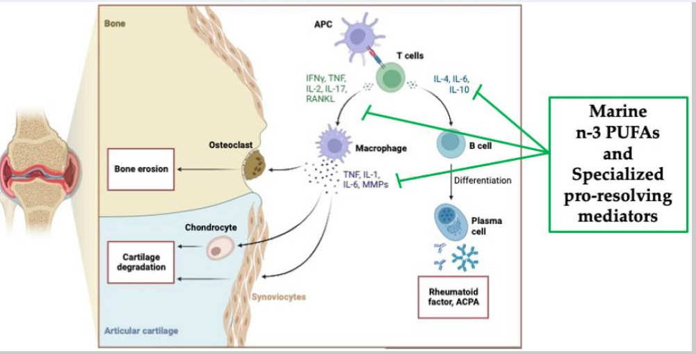
A 2024 review of marine omega-3 PUFA (polyunsaturated fatty acids) for RA reported:

“Altogether the data reported in this review show that anti-inflammatory interventions, i.e., high fish consumption or supplements containing n-3 PUFAs, should be the standard of care, along with pharmacotherapy, in treating patients with RA.”

And here is a graphic from that article showing the effect of SPMs (specialized pro-resolving mediators, derived from omega-3s):

What about osteoarthritis?

A multicenter randomized double blind placebo controlled trial of krill oil containing 0.60 g EPA/d, 0.28 g DHA/d, 0.45 g astaxanthin/d demonstrated improvements in pain, stiffness and physical function.

Some omega-3 supplement studies have demonstrated no significant pain relief in osteoarthritis. Those studies did not reduce the consumption of pro-inflammatory n-6 fatty acids which compete with omega-3 fats for the enzymes which can lead to pro or anti-inflammatory mediators. They also did not measure the omega 6/omega 3 ratio in blood or tissues. Nor did they measure the omega-3 index (% of omega-3 achieved in red blood cell membranes, the gold standard for evaluating tissue levels achieved) This 2018 analysis stated:

“High Omega-3 (n-3) polyunsaturated fatty acids (PUFAs) are associated with lower levels of inflammatory mediators, anti-nociception, and adaptive cognitive/emotional functioning. High Omega-6 (n-6) PUFAs are associated with inflammation, nociception, and psychological distress. While findings related to n-3 supplementation in knee OA are mixed, consideration of the n-6:n-3 ratio and additional outcome measures may provide improved understanding of the potential relevance of these fatty acids in OA”

The authors went on to access blood n-6/n-3 ratios in patients with OA and found the following:

“The high ratio group reported greater pain and functional limitations, (all p’s<0.04), mechanical temporal summation (hand and knee, p<0.05), and perceived stress (p=0.008) but not depressive symptoms.”

“In adults with knee pain, a high n-6:n-3 ratio is associated with greater clinical pain/functional limitations, experimental pain sensitivity, and psychosocial distress compared to a low ratio group.”

The anti-inflammatory diet that I follow and recommend eliminates the major sources of excess omega-6 in the diet, specifically the “vegetable oils” which are actually seed, grain, and legume oils predominated by soy oil, corn oil, peanut and cottonseed oil present in cooking “vegetable oils” and processed foods. A table that displays the ratio of omega 3 to omega 6 in various oils can be found here. Note that this table does not reveal the amounts of MUFA (mono unsaturated fatty acids) which are arguably “heart healthy”. Nor does it address the important issue of protective polyphenols and anti-oxidants (such as in Extra Virgin Olive oil aka EVOO). So do not make choices of oil based only on the omega-3/6 ratio.

Another consideration in choosing oils for cooking (as opposed to salad dressing) is the smoke point. Under high heat, oils are subject to oxidation which creates a proinflammatory effect when consumed. Refined Avocado oil has the highest smoke point (520 degrees F). But we digress. Back to pain and arthritis.

An article just published in Nutrients reviewed Omega-3 Supplementation and Its Effects on Osteoarthritis.

“omega-3 polyunsaturated fatty acids (PUFA) have demonstrated an influential role in the progression of OA, resulting in the reduction of cartilage destruction, inhibition of pro-inflammatory cytokine cascades, and production of oxylipins that promote anti-inflammatory pathways.” 

“Research has demonstrated a positive effect on the modulation of OA symptoms through diet and exercise to promote an anti-inflammatory environment. More specifically, omega-3 PUFAs have demonstrated a reduction in inflammatory biomarkers and cartilage degradation, counteracting the natural disease state of OA. In addition to their chondroprotective role, omega-3 supplementation has been shown to have indirect positive effects on muscle tissue recovery following exercise, which is necessary to prevent the progression of OA and maintain an independent, healthy lifestyle. The effects of omega-3 supplementation on the disease state of OA and its symptoms remain inconclusive. Further clinical trials utilizing human participants are warranted to provide a conclusive recommendation on standardized supplementation of omega-3 for the modulation of osteoarthritis.”

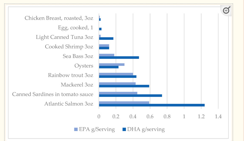
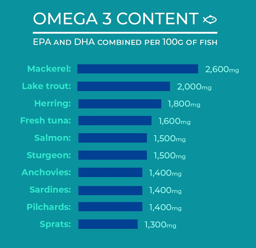
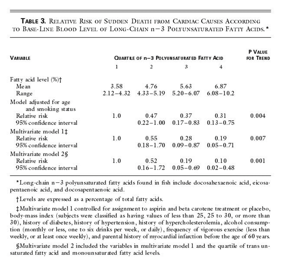
Given the cardioprotective effects, discussed in my last post (including an 80% reduction in sudden death at the highest quintile of omega-3 index) and other benefits (reduction in all cause mortality with high tissue levels), there are many reasons to include large amounts of low mercury fatty fish (wild Alaskan salmon, sardines, herring, trout) in the diet and to consider supplementation when your omega 3 index is < 8%. Likewise, in the presence of arthritis and pain, getting tissue levels of omega 3 up and reducing excessive pro-inflammatory omega 6 will likely provide significant benefit.

Here is a graphic with the omega 3 content of some foods.

And another:

As mentioned in my previous post about omega-3 and cardiovascular health, 1800 mg of omega-3 FA daily is adequate in most people to achieve and omega-3 index of 8%, the level at which cardiovascular protection is greatest.

THIS WEBSITE PROVIDES INFORMATION FOR EDUCATIONAL PURPOSES ONLY. CONSULT YOUR HEALTH CARE PROVIDER FOR MEDICAL ADVICE.

Eat clean, drink filtered water, love, laugh, exercise outdoors in a greenspace, get some morning sunlight, block the blue light before bed, engage in meaningful work, find a sense of purpose, spend time with those you love, AND sleep well tonight.

Doctor Bob

Omega-3 in your diet and supplements

The benefit of omega-3 supplementation has been debated in the cardiology and nutritional literature for many years. Most studies of supplementation have failed to measure tissue levels achieved and often used very low doses. But when tissue levels were measured, either in the serum or red blood cell membrane, the studies consistently demonstrated significant reductions in all-cause mortality and cardiovascular mortality associated with high levels of omega-3 fatty acids.

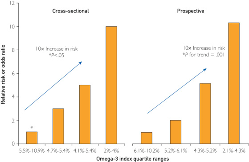
In addition, higher levels of omega 3 are associated with >=80% reduction in sudden death associated with acute myocardial infarction (acute MI) and > 80% reduction in sudden death in cohorts without known coronary artery disease followed long term.

Two Coronary CT Angiogram (CCTA) studies demonstrated that patients with stable coronary artery disease on statin therapy randomized to high dose EPA and DHA had “prevention of coronary plaque progression when an omega-3 fatty acid index >= 4% was achieved.”

 Another CCTA study demonstrated that patients receiving omega 3 supplementation had significantly less coronary atherosclerotic “high risk” lipid rich plaque prevalence (3.8% versus 32%) and lower total non-calcified plaque burden independent of cardiovascular risk factors compared to matched controls not receiving omega 3 supplements.

Omega 3 supplementation after an acute myocardial infarction has been found to reduce infarct size, reduce scaring (fibrosis), and enhance heart tissue healing. (Randomized controlled clinical trial) However a post MI study in 1027 elderly patients randomized to receive 1.8 grams per day of EPA+DHA versus a control group receiving corn oil showed no reduction in the primary composite cardiovascular endpoint between the two groups at 2 years but a higher incidence of AF in the omega 3 group that did not reach statistical significance.

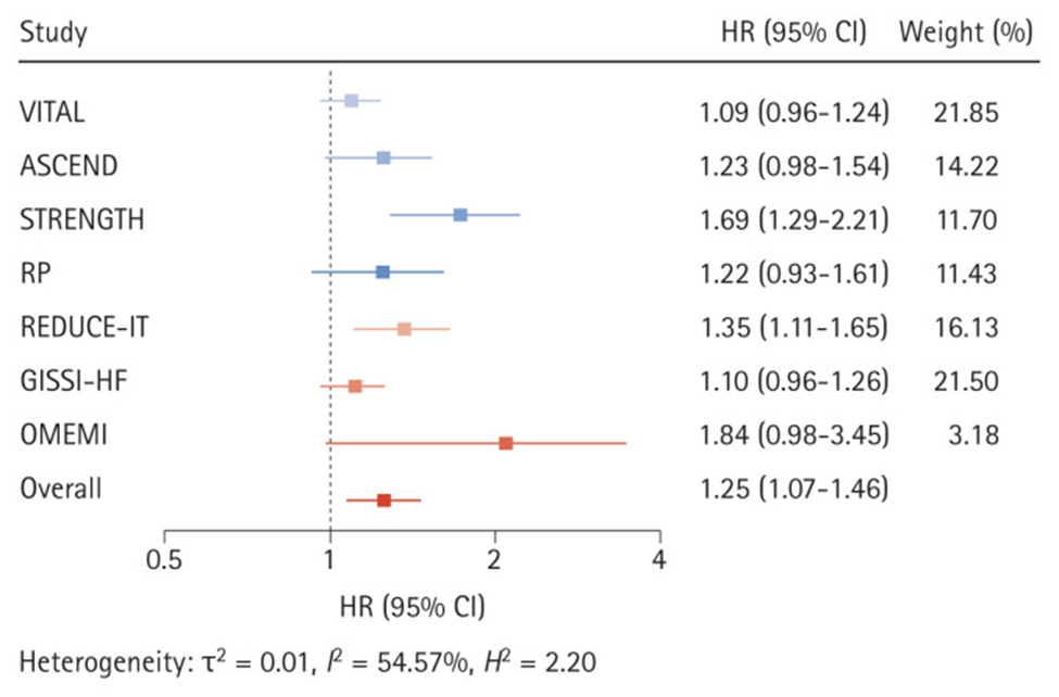
Recently a study, widely reported by the lay press, suggested that high dose omega-3 supplementation was associated with increased risk of atrial fibrillation (AF). These results conflicted with previous studies which demonstrated just the opposite, specifically prior studies demonstrated reduced risk of AF. The more recent study suffered a significant design flaw. The study in question failed to make statistical adjustment for the increased life span associated with higher levels of omega-3. Since age is a primary risk factor for AF, any intervention which increases life span would be expected to result in more AF over the lifetime of the patients as they aged (i.e., more elder years results in increased risk of AF). Therefore, statistical adjustment for that effect should be employed, but was not done in the study.

Unfortunately, science journalism has deteriorated to a state where the conclusions of study authors are most often quoted without interpretation or context, and without critical analysis or comparisons with previous studies that may have demonstrated opposing results.

In addition to large well-designed studies that have suggested a reduced risk of AF associated with omega-3 fatty acids, there have been natural experiments that provide reassuring information. The indigenous Inuit people of Greenland, for example, historically consumed large amounts of omega-3 in their diet with no evidence of increased risk of AF. In fact, before the introduction of western processed foods, estimates of AF among the Inuit were 0.6% (1963) compared to a “worldwide prevalence of AF in adults between 2 and 4%, between one and two percent in Canadian and the general US population and between 0.5% and 3% in most low- and middle-income countries.” A more recent study of Greenland yielded a prevalence of 1.4% likely reflecting a change in habits consisting of less exercise, more tobacco use and a shift to a more Western diet.

Still, multiple studies that used high dose omega 3 supplements in patients with known cardiac disease suggest an increased risk of AF. A good review of omega-3 fatty acids and atrial fibrillation was published in the Korean Journal of Internal Medicine, referenced below.

My interpretation of the complex data in this area is as follows.

At supplemental doses of EPA+DHA above 1.8 grams per day (and perhaps above 1 gram per day) in patients with known coronary artery disease (CAD), at high risk of CAD, or following a myocardial infarction, the risk of AF is increased by about 25% (relative risk). But the risk of lethal ventricular arrythmias (sudden death) associated with myocardial infarction (heart attack) is 80% lower in patients with a red blood cell omega 3 index of >=8. In people without known CAD, an omega-3 index >=8% is associated with an 80% reduction in sudden cardiac death. CCTA studies show significantly lower unstable “vulnerable” plaque in patients on omega-3 supplements. Similarly, omega 3 supplementation in patients on statins associates with halted plaque progression determined by serial CCTA in non-diabetics.

In addition, higher tissue levels of omega 3 are associated with significantly reduced all-cause, cardiovascular, and cancer mortality.

Omega-3 fatty acids are the chemical precursors of SPMs, specialized pro-resolving lipid mediators which help resolve inflammation. We know that cardiovascular events are driven by chronic inflammation in the walls of arteries, often mediated by insulin resistance. Chronic inflammation contributes to atherosclerosis (production of plaque in the artery wall) as well as cardiovascular events that result when unstable plaque ruptures.  Studies suggest that n-3 fatty acids may have antiarrhythmic properties with membrane-stabilizing effects in addition to antithrombotic and anti-inflammatory properties on the endothelial level. Basic science, observational studies and clinical trials have demonstrated that higher tissue levels of omega 3 fatty acids are associated with longer health span and lifespan. This understanding must be balanced with a probable increased risk of AF in certain clinical situations associated with high dose omega-3 supplements as described above (people with known CAD, high risk for CAD, or following and MI). Note that current AHA and ACC dietary guidelines include at least 2 servings of fatty fish per week, one serving provides approximately 1800 mg of omega-3.

Getting omega-3 fatty acids from cold water fatty fish would be ideal. Unfortunately, many individuals do not like salmon, sardines, mackerel or herring and simply will not consume enough of this fish to achieve protective tissue levels. Other species of fish and seafood provide much less amounts of omega 3. Another consideration is that individuals process omega 3 fats differently so different amounts of omega 3 will be necessary to reach the same protective levels in tissue. You can obtain a red blood cell omega-3 index using a home kit and a finger prick without a prescription (https://omegaquant.com/). The sample is mailed in to the lab and results reported directly to you. I have no financial relationship with these folks.

Bill Harris, PhD, is widely published in the area of omega-3 science. He developed the first clinically useful tissue assay which measures the % of omega 3 fat in red blood cell membranes, the “omega-3 index” which is the gold standard for omega 3 research and clinical testing. Although serum levels correlate with the red blood cell index, the later reveals dietary consequences of a 2-3 month period while serum levels reflect just a few days of most recent dietary habits. The red blood cell omega 3 index is analogous to the hemoglobin A1c which reveals average blood sugars over a 2–3-month period. Bill Harris suggests that 1800 mg per day of omega 3 fat consumption (food plus supplements) will achieve an index of >= 8% in most individuals.

Here are some references.

Harris WS, Tintle NL et.al., Fatty Acids and Outcomes Research Consortium (FORCE). Blood n-3 fatty acid levels and total and cause-specific mortality from 17 prospective studies. Nat Communications. 2021 Apr 22;12(1):2329. doi: 10.1038/s41467-021-22370-2. PMID: 33888689; PMCID: PMC8062567. https://pubmed.ncbi.nlm.nih.gov/33888689/

“Here we report the results of a de novo pooled analysis conducted with data from 17 prospective cohort studies examining the associations between blood omega-3 fatty acid levels and risk for all-cause mortality. Over a median of 16 years of follow-up, 15,720 deaths occurred among 42,466 individuals. We found that, after multivariable adjustment for relevant risk factors, risk for death from all causes was significantly lower (by 15-18%, at least p < 0.003) in the highest vs the lowest quintile for circulating long chain (20-22 carbon) omega-3 fatty acids (eicosapentaenoic, docosapentaenoic, and docosahexaenoic acids). Similar relationships were seen for death from cardiovascular disease, cancer and other causes”

Blood Levels of Long-Chain n–3 Fatty Acids and the Risk of Sudden Death Authors: Christine M. Albert, M.D., M.P.H., Hannia Campos, Ph.D., Meir J. Stampfer, M.D., Dr.P.H., Paul M. Ridker, M.D., M.P.H., JoAnn E. Manson, M.D., Dr.P.H., Walter C. Willett, M.D., Dr.P.H., and Jing Ma, M.D., Ph.D.

Published April 11, 2002 N Engl J Med 2002;346:1113-1118DOI:10.1056/NEJMoa012918 VOL. 346 NO. 15 https://www.nejm.org/doi/full/10.1056/NEJMoa012918

We conducted a prospective, nested case–control analysis among apparently healthy men who were followed for up to 17 years in the Physicians’ Health Study. The fatty-acid composition of previously collected blood was analyzed by gas–liquid chromatography for 94 men in whom sudden death occurred as the first manifestation of cardiovascular disease and for 184 controls matched with them for age and smoking status.

RESULTS

Base-line blood levels of long-chain n–3 fatty acids were inversely related to the risk of sudden death both before adjustment for potential confounders (P for trend = 0.004) and after such adjustment (P for trend = 0.007). As compared with men whose blood levels of long-chain n–3 fatty acids were in the lowest quartile, the relative risk of sudden death was significantly lower among men with levels in the third quartile (adjusted relative risk, 0.28; 95 percent confidence interval, 0.09 to 0.87) and the fourth quartile (adjusted relative risk, 0.19; 95 percent confidence interval, 0.05 to 0.71).

CONCLUSIONS

The n–3 fatty acids found in fish are strongly associated with a reduced risk of sudden death among men without evidence of prior cardiovascular disease.

Heydari B, Abdullah S, Pottala JV, Shah R, Abbasi S, Mandry D, Francis SA, Lumish H, Ghoshhajra BB, Hoffmann U, Appelbaum E, Feng JH, Blankstein R, Steigner M, McConnell JP, Harris W, Antman EM, Jerosch-Herold M, Kwong RY. Effect of Omega-3 Acid Ethyl Esters on Left Ventricular Remodeling After Acute Myocardial Infarction: The OMEGA-REMODEL Randomized Clinical Trial. Circulation. 2016 Aug 2;134(5):378-91. doi: 10.1161/CIRCULATIONAHA.115.019949. PMID: 27482002; PMCID: PMC4973577. https://www.ahajournals.org/doi/10.1161/CIRCULATIONAHA.115.019949

Conclusions: Treatment of patients with acute myocardial infarction with high-dose omega-3 fatty acids was associated with reduction of adverse left ventricular remodeling, noninfarct myocardial fibrosis, and serum biomarkers of systemic inflammation beyond current guideline-based standard of care.

Effect of Different Antilipidemic Agents and Diets on Mortality A Systematic Review

Studer M, Briel M, Leimenstoll B, Glass TR, Bucher HC. Effect of Different Antilipidemic Agents and Diets on Mortality: A Systematic Review. Arch Intern Med. 2005;165(7):725–730. doi:10.1001/archinte.165.7.725

Compared with control groups, risk ratios for cardiac mortality indicated benefit from statins (0.78; 95% CI, 0.72-0.84), resins (0.70; 95% CI, 0.50-0.99) and n-3 fatty acids (0.68; 95% CI, 0.52-0.90).

Feuchtner G, Langer C, Barbieri F, Beyer C, Dichtl W, Friedrich G, Schgoer W, Widmann G, Plank F. The effect of omega-3 fatty acids on coronary atherosclerosis quantified by coronary computed tomography angiography. Clin Nutr. 2021 Mar;40(3):1123-1129. doi: 10.1016/j.clnu.2020.07.016. Epub 2020 Jul 22. PMID: 32778459. https://pubmed.ncbi.nlm.nih.gov/32778459/

Conclusions: Omega-3-PUFA supplementation is associated with less coronary atherosclerotic “high-risk” plaque (lipid-rich) and lower total non-calcified plaque burden independent on cardiovascular risk factors. Our study supports direct anti-atherogenic effects of Omega-3-PUFA.

Alfaddagh A, Elajami TK, Saleh M, Mohebali D, Bistrian BR, Welty FK. An omega-3 fatty acid plasma index ≥4% prevents progression of coronary artery plaque in patients with coronary artery disease on statin treatment. Atherosclerosis. 2019 Jun;285:153-162. doi: 10.1016/j.atherosclerosis.2019.04.213. Epub 2019 Apr 13. PMID: 31055222; PMCID: PMC7963401.An omega-3 fatty acid plasma index ≥4% prevents progression of coronary artery plaque in patients with coronary artery disease on statin treatment – PMC (nih.gov)

Conclusions: EPA and DHA added to statins prevented coronary plaque progression in nondiabetic subjects with mean LDL-C <80 mg/dL, when an omega-3 index ≥4% was achieved. Low omega-3 index <3.43% identified nondiabetic subjects at risk of coronary plaque progression despite statin therapy

Association of Plasma Phospholipid Long-Chain Omega-3 FattyAcids with Incident Atrial Fibrillation in Older Adults: The Cardiovascular Health Study, Circulation Volume 125, Number 9 https://doi.org/10.1161/CIRCULATIONAHA.111.062653

Among 3326 US men and women ≥65 years of age and free of AF or heart failure at baseline, plasma phospholipid levels of eicosapentaenoic acid, docosapentaenoic acid, and docosahexaenoic acid were measured at baseline by use of standardized methods. Incident AF (789 cases) was identified prospectively from hospital discharge records and study visit ECGs during 31 169 person-years of follow-up (1992-2006).

Conclusions: In older adults, higher circulating total long-chain n-3 PUFA and docosahexaenoic acid levels were associated with lower risk of incident AF (atrial fibrillation). These results highlight the need to evaluate whether increased dietary intake of these fatty acids could be effective for the primary prevention of AF.

Omega-3 Fatty Acid Therapy: The Tide Turns for a Fish Story https://www.mayoclinicproceedings.org/article/S0025-6196(16)30764-9/fulltext

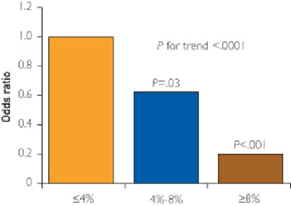
An omega-3 index of less than 4% is associated with increased CHD risk, particularly for sudden cardiac death. In contrast, an omega-3 index of more than 8% is associated with low CHD risk, whereas the range between 4% and 8% is considered intermediate risk

Risk of sudden death

Alfaddagh A, Elajami TK, Ashfaque H, Saleh M, Bistrian BR, Welty FK. Effect of Eicosapentaenoic and Docosahexaenoic Acids Added to Statin Therapy on Coronary Artery Plaque in Patients with Coronary Artery Disease: A Randomized Clinical Trial. J Am Heart Assoc. 2017; 6: e006981. 10.1161/JAHA.117.006981. https://pubmed.ncbi.nlm.nih.gov/29246960/

“High-dose eicosapentaenoic acid and docosahexaenoic acid provided additional benefit to statins in preventing progression of fibrous coronary plaque in subjects adherent to therapy with well-controlled low-density lipoprotein cholesterol levels.”

Huh JH, Jo SH. Omega-3 fatty acids and atrial fibrillation. Korean J Intern Med. 2023 May;38(3):282-289. doi: 10.3904/kjim.2022.266. Epub 2022 Dec 14. PMID: 36514212; PMCID: PMC10175873 https://pubmed.ncbi.nlm.nih.gov/36514212/

.

Effects of omega-3 fatty acid supplementation on the risk of atrial fibrillation. HR, hazard ratio; CI, confidence interval; VITAL, Vitamin D and Omega-3 Trial; ASCEND, A Study of Cardiovascular Events in Diabetes; STRENGTH, Statin Residual Risk with Epanova in High Cardiovascular Risk Patients with Hypertriglyceridemia; RP, Risk and Prevention Study; REDUCE-IT, Reduction of Cardiovascular Events With Icosapent Ethyl-Intervention Trial; GISSI-HF, Gruppo Italiano per lo Studio della Sopravvivenza nell’Insufficienza Cardiaca-Heart Failure; OMEMI, Omega-3 Fatty Acids in Elderly With Myocardial Infarction. Effects of omega-3 fatty acid supplementation on the risk of atrial fibrillation. HR, hazard ratio; CI, confidence interval; VITAL, Vitamin D and Omega-3 Trial; ASCEND, A Study of Cardiovascular Events in Diabetes; STRENGTH, Statin Residual Risk with Epanova in High Cardiovascular Risk Patients with Hypertriglyceridemia; RP, Risk and Prevention Study; REDUCE-IT, Reduction of Cardiovascular Events With Icosapent Ethyl-Intervention Trial; GISSI-HF, Gruppo Italiano per lo Studio della Sopravvivenza nell’Insufficienza Cardiaca-Heart Failure; OMEMI, Omega-3 Fatty Acids in Elderly With Myocardial Infarction. Effects of omega-3 fatty acid supplementation on the risk of atrial fibrillation. HR, hazard ratio; CI, confidence interval; VITAL, Vitamin D and Omega-3 Trial; ASCEND, A Study of Cardiovascular Events in Diabetes; STRENGTH, Statin Residual Risk with Epanova in High Cardiovascular Risk Patients with Hypertriglyceridemia; RP, Risk and Prevention Study; REDUCE-IT, Reduction of Cardiovascular Events With Icosapent Ethyl-Intervention Trial; GISSI-HF, Gruppo Italiano per lo Studio della Sopravvivenza nell’Insufficienza Cardiaca-Heart Failure; OMEMI, Omega-3 Fatty Acids in Elderly With Myocardial Infarction

THIS WEBSITE PROVIDES INFORMATION FOR EDUCATIONAL PURPOSES ONLY. CONSULT YOUR HEALTH CARE PROVIDER FOR MEDICAL ADVICE.

Eat clean, drink filtered water, love, laugh, exercise outdoors in a greenspace, get some morning sunlight, block the blue light before bed, engage in meaningful work, find a sense of purpose, spend time with those you love, AND sleep well tonight.

Doctor Bob

4th International Evolutionary Health Conference

Sorry for the confusion. The website for the International Evolutionary Health Conference changed when the venue changed from Boston to Virtual. Here is the correct website link which gives a list of speakers/topics and sign up information. 

https://2023.evolutionaryhealthconference.com/

The previously published link will lead you to a site that says “canceled”. The conference is not cancelled, the venue has changed to virtual. 

Dr. Bob

/

Fourth International Evolutionary Health Conference

I’ve been asked to talk at the fourth International Evolutionary Health Conference on the topic of Cardiovascular Risk Assessment. This year the conference is virtual. Presenters include clinicians and researchers discussing many topics related to health. The underlying principle of this approach attributes modern degenerative and chronic diseases to mismatch between our evolutionary biology and present day life. You can sign up for this virtual event here.

https://2023.evolutionaryhealthconference.com/

Agenda

9:45 AM – 10:00 AM

Opening remarks

Prof. Lynda Frassetto

10:00 AM – 10:30 AM

Maladaptive cognitive/emotional processing as the cause of the stress response

Prof. Igor Mitrovic


Physiologic reserve is spare capacity activated when demand exceeds baseline, causing stress. If demand surpasses reserve, it damages the system and leads to death. The brain predicts the future to a…
View More

10:30 AM – 11:00 AM

How breathing patterns affect health

Dr. Michael Mew

11:00 AM – 11:15 AM

Round table with Q & A (Moderator: Darryl Edwards)

Dr. Michael Mew

Prof. Igor Mitrovic

11:15 AM – 11:45 AM

Break and Poster session


If you would like to submit a poster, please contact us at evolution.conference@nutriscience.pt

11:45 AM – 12:15 PM

Decoding The Truth: Cancer, Carbs and Cure

Darryl Edwards, MSc


1. We will delve into the extensive evidence showcasing how higher levels of physical activity can reduce the risk of various cancers. 2. While awareness of the importance of exercise exists, we will…
View More

12:15 PM – 12:45 PM

Influential factors on sun-induced vitamin D synthesis

Pedro Bastos, PhD candidate


Ultraviolet B radiation is absorbed in the epidermis by 7-dehydrocholesterol, giving rise to previtamin D3 and subsequently to vitamin D3. In the liver, vitamin D is converted to one of the various c…
View More

12:45 PM – 1:00 PM

Round table with Q & A (Moderator: Prof. Lynda Frassetto)

Darryl Edwards, MSc

Pedro Bastos, PhD candidate

1:00 PM – 2:15 PM

Lunch Break

2:15 PM – 2:45 PM

How nutrition can impact microbiome composition/permeability/immune response

Prof. Alessio Fasano


Improved hygiene and reduced microorganism exposure are linked to the ‘epidemic’ of chronic inflammatory diseases (CID) in developed nations. This hygiene hypothesis suggests that lifestyle and envir…
View More

2:45 PM – 3:15 PM

Comprehensive cardiovascular risk assessment

Dr. Robert Hansen


Assessing insulin resistance is central to predicting CV risk. LDL-C and standard lipid profile is extremely limited in predictive value. A systems engineering understanding of atherosclerosis and ev…
View More

3:15 PM – 3:30 PM

Round table with Q & A (Moderator: Pedro Bastos)

Prof. Alessio Fasano

Dr. Robert Hansen

3:30 PM – 3:45 PM

Short Break

3:45 PM – 4:15 PM

Environmental influences on cellular senescence and aging

Prof. Peter Stenvinkel


Planetary health recognizes that human well-being depends on the health of ecosystems. Neglecting this concept has led to an anthropocentric world, causing increased greenhouse gas emissions, heat st…
View More

4:15 PM – 4:45 PM

Fueling a Bright Future: The Role of Diet in Preventing Childhood Obesity

Dr. Polina Sayess


Childhood obesity is a global health issue. In my presentation, I’ll explore its origins, classifications, and mitigation strategies. I’ll discuss the definitions and distinctions between “overweight…
View More

4:45 PM – 5:00 PM

Round table with Q & A (Moderator: Prof. Lynda Frassetto)

Prof. Peter Stenvinkel

Dr. Polina Sayess

5:00 PM – 5:30 PM

Final discussion with all speakers and moderators


Establishing future research and intervention directions.

5:30 PM – 5:45 PM

Closing remarks

Prof. Lynda Frassetto

Please join us if you can.

Dr. Bob

Long Haulers, Brain Fog and Post Exertional Malaise

An excellent article recently published in the Atlantic was so well written that I have cut and pasted important snippets to help create this post. The review confirms many findings uncovered in my reading of several scientific publications.

You can read the full article here:

The crushing fog of long COVID

20 to 30 percent of patients report brain fog three months after their initial infection, as do 65 to 85 percent of the long-haulers who stay sick for much longer.

Of long COVID’s many possible symptoms, brain fog “is by far one of the most disabling and destructive,”

It is more profound than the clouded thinking that accompanies hangovers, stress, or fatigue.

It is not a mood disorder.

It is almost always a disorder of “executive function”—the set of mental abilities that includes:

  1. focusing attention,
  2. holding information in mind, and
  3. blocking out distractions.

Patients state they often lose focus mid-sentence.

Difficulty with simple tasks impairs activities of daily living.

“I couldn’t unload a dishwasher, because identifying an object, remembering where it should go, and putting it there was too complicated.”

The memories are there, but with impaired executive function, the brain neither chooses the important things to store nor retrieves that information efficiently.

Most people with brain fog are not so severely affected, and gradually improve with time. But even when people recover enough to work, they can struggle with minds that are less nimble than before.

“I’ve had surgeons who can’t go back to surgery, because they need their executive function,” Monica Verduzco-Gutierrez, a rehabilitation specialist at UT Health San Antonio.

That specific constellation of problems also befalls many people living with HIV, epileptics after seizures, cancer patients experiencing so-called chemo brain, and people with several complex chronic illnesses such as fibromyalgia.

It’s part of the diagnostic criteria for myalgic encephalomyelitis, also known as chronic fatigue syndrome, or ME/CFS

People with brain fog also excel at hiding it: to protect their jobs when still able to work, or to protect their reputation, or out of embarrassment.

“I know my value in many people’s eyes will be diminished by knowing that I have these cognitive challenges.”

Individuals with previously above average cognitive ability often test “normal” but suffer significant loss compared to their prior ability.

A team of British researchers analyzed data from the UK Biobank study. The findings revealed structural changes in the brain with loss of tissue on MRI scans that correlates with symptoms.

They found that even mild infections can slightly shrink the brain and reduce the thickness of its neuron-rich gray matter. At their worst, these changes were comparable to a decade of aging.

They were especially pronounced in areas such as the parahippocampal gyrus, which is important for encoding and retrieving memories, and the orbitofrontal cortex, which is important for executive function.

In most cases the virus probably harms the brain without directly infecting it.

Inflammatory chemicals can travel from the lungs to the brain, where they disrupt cells called microglia (immune cells in the brain).

In their presence, the hippocampusa region crucial for memoryproduces fewer fresh neurons, while many existing neurons lose their insulating coats (demyelination), so electric signals now course along these cells more slowly.

These are the same changes seen in cancer patients with “chemo fog”.

Neuro-inflammation is “probably the most common way” that COVID results in brain fog, but that there are likely many such routes, such as reactivation of dormant viruses such as Epstein-Barr virus, which has been linked to conditions including ME/CFS and multiple sclerosis.

By damaging blood vessels and filling them with small clots, COVID also throttles the brain’s blood supply, depriving this most energetically demanding of organs of oxygen and fuel.

These problems can be exacerbated or mitigated by factors such as sleep and rest, which explains why many people with brain fog have good days and bad days.

Although other respiratory viruses can wreak inflammatory havoc on the brain, SARS-CoV-2 does so more potently than influenza.

For adults following SARS CoV-2 infection:

risks of cognitive deficit (known as brain fog), dementia, psychotic disorders, and epilepsy or seizures were still increased at the end of the 2-year follow-up period.

For children following SARS CoV-2 infection:

in the 6 months after SARS-CoV-2 infection, children were not at an increased risk of mood (HR 1·02 [95% CI 0·94–1·10) or anxiety (1·00 [0·94–1·06]) disorders, but did have an increased risk of cognitive deficit, insomnia, intracranial haemorrhage, ischaemic stroke, nerve, nerve root, and plexus disorders, psychotic disorders, and epilepsy or seizures (HRs ranging from 1·20 [1·09–1·33] to 2·16 [1·46–3·19])….  Unlike adults, cognitive deficit in children had a finite risk horizon (75 days) and a finite time to equal incidence (491 days).

The fact that neurological and psychiatric outcomes were similar during the delta and omicron waves indicates that the burden on the health-care system might continue even with variants that are less severe in other respects.

There are no proven drug treatments for long-haulers.

But there is hope.

Cancer researchers have developed drugs that can calm inflamed microglia in mice and restore their cognitive abilities;

“Metformin can promote the regeneration of neural precursor cell populations and improve cognitive function in a preclinical model of cranial radiation and a pilot clinical study of children after cranial radiation and chemotherapy.”

Treating cancer therapy–related cognitive impairment | Nature Medicine

With regard to long-haulers, better sleep, healthy eating, and other generic lifestyle changes can make the condition more tolerable. Breathing and relaxation techniques can help people through bad flare-ups; speech therapy can help those with problems finding words.

“Some people spontaneously recover back to baseline,”

The largest group of long-haulers—those whose brain fog has improved but not vanished, can “maintain a relatively normal life, but only after making serious accommodations,”

Patients struggle to make peace with how much they’ve changed and the stigma associated with it, regardless of where they end up.

Post-exertional malaise—severe crashes in which all symptoms worsen after even minor physical or mental exertion is commonly reported.

Many long-haulers try to push themselves back to work and instead “push themselves into a crash,”

Post-exertional malaise is so common among long-haulers that “exercise as a treatment is inappropriate for people with long COVID,”

Even brain-training games—which have questionable value but are often mentioned as potential treatments for brain fog—must be very carefully rationed because mental exertion is physical exertion.

People with ME/CFS learned this lesson the hard way, and fought hard to get exercise therapy, once commonly prescribed for the condition, to be removed from official guidance in the U.S. and U.K.

In summary:

  1. Brain fog can occur even after mild or asymptomatic Covid-19.
  2. Although many patients improve over time, many are left with disability that can range from mild to incapacitating.
  3. Although these symptoms can occur following any viral infection, SARS CoV-2 seems to produce this with greater frequency compared to other viruses.
  4. Chronic brain inflammation is the likely cause in many individuals.
  5. Reactivation of Epstein Barr and/or other dormant viruses is suggested by various immune markers.
  6. The immune signature also suggests an immune response that mimics persistent infection in the absence of live SARS CoV-2 virus.
  7. Post exertional malaise following physical or mental exercise is a common and debilitating symptom without proven treatments. However there are guidelines that may help mitigate this devastating condition.
  8. Brain fog and post-exertional malaise are hallmarks of chronic fatigue syndrome/Myalgic encephalomyelitis which can occur following viral infections and major stress events such as physical and psychological trauma.
  9. Although not discussed in this post, chronically suppressed cortisol levels have been identified in this population. This in combination with physical changes noted on brain MRI demonstrate that there are physical correlates of brain fog.

In the meantime, preventive measures represent the low hanging fruit for health in general and with respect to the pandemic.

In the context of the COVID 19 pandemic I will close with the usual summary.

  1. Avoid alcohol consumption (alcohol wreaks havoc with your immunity)
  2. Get plenty of sleep (without adequate sleep your immune system does not work well )
  3. Follow good sleep habits
  4. Exercise, especially out of doors in a green space, supports the immune system
  5. Get some sunshine and make sure you have adequate Vitamin D levels. Supplement with Vitamin D3 to get your levels above 30 ng/ml, >40ng/ml arguably better.
  6. Eat an anti-inflammatory diet rich in micronutrients.
  7. Practice stress reduction like meditation and yoga which improves the immune system
  8. Eliminate sugar-added foods and beverages from your diet. These increase inflammation, cause metabolic dysfunction, and suppress immunity.
  9. Eliminate refined-inflammatory “vegetable oils” from your diet, instead eat healthy fat.
  10. Clean up your home environment and minimize your family’s exposure to environmental toxins by following recommendations at EWG.org with regards to household products, personal care products, and organic foods. (https://www.ewg.org/)
  11. Drink water filtered through a high quality system that eliminates most environmental toxins.
  12. If you are eligible for vaccination, consider protecting yourself and your neighbor with a few jabs. Age > 50 and/or risk factors means clear benefit from a booster.

THIS WEBSITE PROVIDES INFORMATION FOR EDUCATIONAL PURPOSES ONLY. CONSULT YOUR HEALTH CARE PROVIDER FOR MEDICAL ADVICE.

Eat clean, drink filtered water, love, laugh, exercise outdoors in a greenspace, get some morning sunlight, block the blue light before bed, engage in meaningful work, find a sense of purpose, spend time with those you love, AND sleep well tonight.

Doctor Bob

AHS 2022 Lecture, Acute and Long Covid, Nutritional and Lifestyle Immunology

I recently gave a talk at the AHS 2022 meeting held at UCLA. You can view the video here:

This first slide gives a good overview.

The presentation covers a quick review of my presentations given last year at the PAH 2021 annual meeting (virtual) with additional information on long Covid.

Multiple nutrients acting synergistically support a balanced response to viral infections, including SARS CoV-2. Here is a picture.

The take home message is that no single nutritional intervention is likely to have significant impact with an acute infection unless all but one nutritional component is optimal. Nevertheless, there is compelling evidence that Vitamin D deficiency is rampant in the developed world and if one nutritional intervention is likely to be of benefit, Vitamin D supplementation, particularly in high risk populations, presents the most likely candidate. As usual, preventive supplementation would be preferable to rescue high dose intervention.

In a study of frail elderly hospitalized patients, regular vitamin D supplementation was associated with decreased mortality as demonstrated here. Compared to no supplementation, regular supplementation was associated with a 93% reduction in risk of death.

A study from Spain with very high dose Vitamin D in the form of Calcifediol showed significant benefit in hospitalized patients, suggesting that Vitamin D deficiency was prevalent in that population and that such a treatment intervention should be widely considered.

Calcifediol Treatment and COVID-19-Related Outcomes

The following graphic from another nutrition review article, with red additions added by myself, demonstrates the complex interaction between nutrition and the two main components of our immune system, innate immunity (immediate response) and adaptive immunity (based on immune memory). Again red highlights added by yours truely.

And here is a slide from my lecture with quotes from that article.

Yet most Americans are deficient in many of these essential nutrients as depicted here. The percentages represent the % of Americans that fall below the estimated amount required to prevent deficiency in HALF of adults (a very low standard).

The EAR is a very low bar to meet, yet many Americans fall below even that low standard.

The SARS CoV2 virus interferes with a crucial component of the the initial (innate) immune response, the production of interferon 1 and the signaling of interferon one to immune cell mediators as depicted in this graphic.

SARS CoV2 on the left is compared to Virus X on the right. On the left interferon 1 (IFN) production and signaling is blocked by the virus, interfering with an effective and controlled immune response, on the right IFN is not blocked. A cascade of events results in TOO LITTLE, TOO LATE, AND THEN TOO MUCH of an immune response, producing a cytokine storm.

Obesity, insulin and leptin resistance, also interfere with the production and signaling of interferon. The result is that people with insulin and leptin resistance (pre-diabetes, Type 2 Diabetes as well as sarcopenia) experience a double hit. First the virus itself disrupts the immune response and superimposed upon the viral effect is the effect of insulin and leptin resistance on the immune response.

SOCS: suppressor of cytokine signaling. Several recent viral studies have shown that viral genes can hijack SOCS1 to inhibit host antiviral pathways, as a strategy to evade host immunity
On the left Interferon production and signaling are normal and a successful immune response is mounted. On the right the presence of insulin and leptin resistance, associated with obesity results in an initial inadequate response and a late excessive response. TOO LITTLE, TOO LATE, THEN TOO MUCH.

Factors that can quickly impact insulin and leptin resistance include all the components of an ancestral lifestyle depicted in my website graphic. A paleolithic or ancestral diet that eliminates sugar added foods and beverages, replacing those empty calories with nutrient dense foods, exercise, adequate restorative sleep, stress reduction, avoidance of environmental toxins, social connection. All of these affect health in general, mitigate insulin and leptin resistance, and support a balanced immune response to viral infection. The circle of health depicted below is surrounded by the many deleterious aspects of modern living. Thus, a mismatch between our evolutionary biology and present day life.

Here is a slide from my lecture that lists many lifestyle factors that can impact infection with any virus, including SARS CoV-2

My lecture also included discussion of Long COVID, theories of etiology and pathophysiology which will be discussed in my next post.

For the full lecture which is about 34 minutes long, please follow the link above.

THIS WEBSITE PROVIDES INFORMATION FOR EDUCATIONAL PURPOSES ONLY. CONSULT YOUR HEALTH CARE PROVIDER FOR MEDICAL ADVICE.

Eat clean, drink filtered water, love, laugh, exercise outdoors in a greenspace, get some morning sunlight, block the blue light before bed, engage in meaningful work, find a sense of purpose, spend time with those you love, AND sleep well tonight.

Doctor Bob

Long Covid: Vascular Inflammatory Response Mediated by non-classical monocytes.

Today I listened to Dr. Bruce Patterson discuss his research on Long-Covid.

Bruce Patterson was director of a virology lab at Stanford before establishing Incelldx, a biotech firm in the area of virus diagnostics. Bruce was interviewed today (second half of the program) on The People’s Pharmacy (NPR). The transcript and podcast will be available tomorrow at PeoplesPharmacy.com (show #1273)

Utilizing AI and data from over 10,000 patients, Incelldx has developed diagnostic tools to characterize the immune system dysregulation associated with long covid. According to Dr. Patterson’s research, Long-Covid involves “non-classical monocytes” that are reacting to remnant virus proteins (not RNA or DNA), producing a vascular inflammatory process. That was the missing link. Researchers were looking for RNA, but the problem appears to be in the monocyte reaction to remnant viral proteins that stimulate these specialized monocytes, producing a chronic vasculitis.

Here is a link to the Long Covid clinical program based upon this research.

https://covidlonghaulers.com/

The program involves immune testing by Incelldx and based on the results, treatment recommendations are made.

So far 2 drugs repurposed for long-Covid appear effective when used in combination.

A CCR5 antagonist (maraviroc) which has been used to treat HIV and a Statin medication which blocks binding of the monocytes to artery walls.

According to Dr. Patterson, Long Covid in many patients is a vascular inflammatory process, mediated by non-classical monocytes which are activated by viral remnant proteins.

Dr. Patterson has seen many patients respond to this drug combination. He uses an old, early statin drug (pravastatin) which has a low side effect profile compared to the more commonly used atorvastatin (which gave me severe myopathy, notorious for that problem but understated by drug company reports).

This is cutting edge, most physicians, even in university settings, are not aware of this diagnostic/therapeutic approach.

This is still considered experimental but appears to be very promising.

In the context of the COVID 19 pandemic I will close with the usual summary.

  1. Avoid alcohol consumption (alcohol wreaks havoc with your immunity)
  2. Get plenty of sleep (without adequate sleep your immune system does not work well )
  3. Follow good sleep habits
  4. Exercise, especially out of doors in a green space, supports the immune system
  5. Get some sunshine and make sure you have adequate Vitamin D levels. Supplement with Vitamin D3 to get your levels above 30 ng/ml. (read this Open Letter)
  6. Eat an anti-inflammatory diet rich in micronutrients.
  7. Practice stress reduction like meditation and yoga which improves the immune system
  8. Eliminate sugar-added foods and beverages from your diet. These increase inflammation, cause metabolic dysfunction, and suppress immunity.
  9. Eliminate refined-inflammatory “vegetable oils” from your diet, instead eat healthy fat.
  10. Clean up your home environment and minimize your family’s exposure to environmental toxins by following recommendations at EWG.org with regards to household products, personal care products, and organic foods. (https://www.ewg.org/)
  11. If you are over age 12 and eligible for vaccination, consider protecting yourself and your neighbor with vaccination.

THIS WEBSITE PROVIDES INFORMATION FOR EDUCATIONAL PURPOSES ONLY. CONSULT YOUR HEALTH CARE PROVIDER FOR MEDICAL ADVICE.

Eat clean, drink filtered water, love, laugh, exercise outdoors in a greenspace, get some morning sunlight, block the blue light before bed, engage in meaningful work, find a sense of purpose, spend time with those you love, AND sleep well tonight.

Doctor Bob