Tag Archives: wellness

Can a drug replace the benefits of exercise?

A new drug, still under investigation and not yet FDA approved is reported to provide the benefits of exercise in humans. The drug is called ATX304 and has been studied in humans in phase II clinical trials. It works by decreasing the degradation of a master energy controller called AMPK (AMP-activated protein kinase) By decreasing the degradation, it increases the amount of AMPK which has multiple favorable metabolic effects. When we exercise our muscles utilize ATP (adenosine triphosphate) the major energy source for our cells. When ATP is broken down the levels of AMP (adenosine monophosphate) increase and that increase signals AMPK to increase. ATX304 produces the same rise in AMPK experienced with exercise. The result is that muscle cells uptake glucose from the circulation without the need for insulin (non insulin dependent glucose uptake), the same occurs with exercise. With exercise the effect can last for 16 hours. Thus blood sugar is better controlled with both exercise and with ATX304. Along with better blood sugar control, insulin sensitivity is maintained and improved by avoiding pancreatic beta cell stress (beta cells produce insulin). ATX-304 also improves blood vessel flow, cardiac pump function, and as a result improved delivery of oxygen and nutrients to muscle and all parts of the body.

ATX304 also increases the uptake of fatty acids by the mitochondria (the organelle in our cells that produce energy in the form of ATP), facilitating energy production through the burning of fat (a process called beta oxidation). This happens without the muscle loss associated with GLP-1 drugs. Muscle is protected because glucose and fatty acid delivery to muscles are enhanced while keeping cellular ATP stable, completely bypassing the starvation pathways that cause muscle wasting when people lose weight by fasting or taking GLP-1 agonist drugs.

While I do not advocate taking pills instead of exercise I view ATX-304 as a better alternative weight loss medication compared to GLP-1 drugs for several reasons. First and foremost one can avoid the 20-40% loss of muscle associated with GLP-1 drugs. Secondly, ATX-304 increases metabolic rate, causing greater energy expenditure.

While ATX-304 represents an exciting potential approach to weight loss while improving insulin sensitivity without loss of muscle, you cannot build muscle strength, muscle mass, improve balance and coordination, or challenge your brain (with the resulting cognitive protection) by taking a pill. You must exercise including resistance training to accomplish those specific beneficial outcomes.

For a deeper dive into ATX-304 and a more detailed understanding how our bodies use glucose and fat to produce energy, as well as understanding how exercise works to improve metabolic health, what follows is a conversation I had with Google Gemini.

Conversation with Gemini

how does atx304 improve glucose control, enhance exercise performance and reduce body fat while maintaining muscle, use only peer reviewed scientific papers and cite references

ATX-304 (formerly known as O304) is a first-in-class, small-molecule, non-allosteric PAN-AMPK activator (AMP-activated protein kinase) currently in clinical development for metabolic and cardiovascular diseases (Ericsson et al., 2021; Steneberg et al., 2018). It functions primarily by protecting phosphorylated AMPK (p-AMPK) from dephosphorylation by protein phosphatases like PP2C, effectively acting as an “exercise mimetic” that triggers catabolic, energy-generating pathways (Steneberg et al., 2018).

Peer-reviewed academic research details the distinct physiological mechanisms through which ATX-304 improves glucose control, enhances exercise performance, and selectively targets body fat while preserving lean muscle mass:

1. Improvement of Glucose Control

ATX-304 manages blood sugar levels through a dual mechanism targeting both skeletal muscle and pancreatic tissues:

  • Insulin-Independent Glucose Uptake: In skeletal muscle, ATX-304 activates AMPK signaling, which drives the translocation of glucose transporter 4 (GLUT4) vesicles to the cell membrane. This allows muscle tissue to pull glucose from the bloodstream independently of insulin action (Steneberg et al., 2018).
  • Beta-Cell Preservation and Rest: By reducing systemic glucose demand and chronic hyperglycemia, the compound mitigates pancreatic β-cell stress. Clinical evidence from Phase IIa trials in patients with Type 2 Diabetes (T2D) demonstrated substantial reductions in fasting plasma glucose (FPG) and overall insulin resistance, quantified by HOMA-IR (Steneberg et al., 2018).
  • Reversal of Age-Related Insulin Resistance: In longitudinal animal models, prolonged administration of the compound prevented the progressive, age-associated hyperinsulinemia that typically induces metabolic syndrome (Ericsson et al., 2021).

2. Enhancement of Exercise Performance

As an exercise mimetic, ATX-304 induces cellular adaptations similar to endurance training:

  • Cardiovascular & Microvascular Remodeling: ATX-304 mimics the cardiac stressors of exercise by enhancing left ventricular stroke volume, increasing end-diastolic volume, and lowering resting heart rate without inducing pathological cardiac hypertrophy (Ericsson et al., 2021; Steneberg et al., 2018). Furthermore, it improves peripheral microvascular perfusion and blood pressure regulations.
  • Mitochondrial Homeostasis: The compound enhances mitochondrial respiration, upregulates fatty acid oxidation, and increases baseline oxygen consumption rates (OCR) in metabolic tissues (Katerelos et al., 2024). These changes directly translate to significantly improved exercise capacity and total work endurance, especially in aged or metabolically compromised subjects (Ericsson et al., 2021).

3. Reduction of Body Fat While Maintaining Muscle

One of the key advantages of direct pan-AMPK activation via ATX-304 is its ability to execute a systemic “metabolic switch” that differentiates between fat tissue and lean body mass:

  • Upregulation of Lipid Oxidation: ATX-304 stimulates cellular catabolism by phosphorylating Acetyl-CoA Carboxylase (ACC). Phosphorylation inhibits ACC, which decreases malonyl-CoA levels, disinhibits carnitine palmitoyltransferase-1 (CPT1), and permits rapid fatty acid oxidation in both the liver and adipose tissue (Holm et al., 2024).
  • Selective Adiposity Loss: Body composition analysis via EchoMRI reveals that the rapid weight reduction induced by ATX-304 corresponds almost entirely to a loss in adipose (fat) mass (Holm et al., 2024). It also limits hepatic de novo lipogenesis and drives down blood cholesterol levels.
  • Muscle Preservation (Lean Weight Maintenance): Unlike severe caloric restriction or certain anti-obesity medications that trigger concurrent muscle loss, animals treated with ATX-304 maintain their absolute lean weight (muscle mass) (Holm et al., 2024). The compound drives a significant long-term increase in energy expenditure, meaning that even when food intake naturally rebounds or increases by 20% to 30%, fat mass continues to decrease while skeletal muscle architecture is protected (Holm et al., 2024).

References

Ericsson, M., Steneberg, P., Nyrén, R., & Edlund, H. (2021). AMPK activator O304 improves metabolic and cardiac function, and exercise capacity in aged mice. Communications Biology, 4(1), 1–12. https://doi.org/10.1038/s42003-021-02837-0 Cited by: 30

Holm, E., Vermeulen, I., Parween, S., López-Pérez, A., Cillero-Pastor, B., Vandenbosch, M., Remeseiro, S., & Hörnblad, A. (2024). AMPK-activator ATX-304 reduces oxidative stress and improves MASLD via metabolic switching. JCI Insight, 9(7). https://doi.org/10.1101/2024.02.13.578901 Cited by: 10

Katerelos, M., Gleich, K., Harley, G., Loh, K., Oakhill, J. S., Kemp, B. E., de Souza, D. P., Narayana, V. K., Coughlan, M. T., Laskowski, A., Ling, N. X. Y., Murray-Segal, L., Brink, R., Lee, M., Power, D. A., & Mount, P. F. (2024). The AMPK activator ATX-304 alters cellular metabolism to protect against cisplatin-induced acute kidney injury. Biomedicine & Pharmacotherapy, 175, 116730. https://doi.org/10.1016/j.biopha.2024.116730 Cited by: 9

Steneberg, P., Lindahl, E., Dahl, U., Lidh, E., Straseviciene, J., Backlund, F., Kjellkvist, E., Berggren, E., Lundberg, I., Bergqvist, I., Ericsson, M., Eriksson, B., Linde, K., Westman, J., Edlund, T., & Edlund, H. (2018). PAN-AMPK activator O304 improves glucose homeostasis and microvascular perfusion in mice and type 2 diabetes patients. JCI Insight, 3(12). https://doi.org/10.1172/jci.insight.99114 Cited by: 132

how do the effects on ACC and CPT1 lead to increased fat oxidation

The biochemical mechanism linking Acetyl-CoA Carboxylase (ACC) and Carnitine Palmitoyltransferase-1 (CPT1) is the primary pathway through which AMPK activators like ATX-304 accelerate fat oxidation.

In cellular metabolism, CPT1 is the “gatekeeper” for fat burning, but its activity is strictly regulated by a molecular brake called malonyl-CoA. Direct activation of AMPK breaks this cycle, shifting the cell into a dedicated fat-burning state.

The Molecular Chain Reaction

When ATX-304 activates AMPK, it initiates a sequential cascade that directly impacts the mitochondria:

        ATX-304 / AMPK Activated

                   │

                   ▼

         ACC is Phosphorylated

             (Inactivated)

                   │

                   ▼

       Malonyl-CoA Levels Drop

         (The “Brake” is Removed)

                   │

                   ▼

        CPT1 is Disinhibited

             (Gate Opens)

                   │

                   ▼

 Fatty Acids Enter Mitochondria ──► Beta-Oxidation (Fat Burning)

1. The Inactivation of ACC

Under normal conditions, the enzyme ACC converts acetyl-CoA into malonyl-CoA. Malonyl-CoA is the fundamental building block for synthesizing new fatty acids (de novo lipogenesis).

When ATX-304 activates AMPK, AMPK attaches a phosphate group to ACC (specifically the ACC1 and ACC2 isoforms). This phosphorylation inactivates ACC, halting the production of malonyl-CoA.

2. The Disinhibition of CPT1

Malonyl-CoA acts as a powerful allosteric inhibitor (a biological “off switch”) for CPT1. CPT1 is an enzyme anchored to the outer mitochondrial membrane. Long-chain fatty acids cannot cross into the mitochondria on their own; they must be attached to carnitine by CPT1 to gain entry.

Because ATX-304 shuts down ACC, cellular levels of malonyl-CoA rapidly drop. Without malonyl-CoA around to inhibit it, CPT1 transitions into its fully active state.

3. Increased Mitochondrial Beta-Oxidation

With the CPT1 gateway open, long-chain fatty acids are rapidly converted into acyl-carnitines, allowing them to pass through the mitochondrial membranes via the carnitine shuttle. Once inside the mitochondrial matrix, these fatty acids undergo β-oxidation (beta-oxidation).

During β-oxidation, the fatty acids are chopped down into acetyl-CoA molecules, which directly feed into the Tricarboxylic Acid (TCA) cycle to generate ATP (cellular energy).

Summary: By turning off ACC, ATX-304 depletes the molecule (malonyl-CoA) that normally blocks CPT1. This unlocks the mitochondrial gateway, forcing the cell to pull circulating fatty acids out of storage and burn them for fuel, ultimately driving down systemic body fat (Holm et al., 2024; Steneberg et al., 2018).

Want to know how this specific fat-burning pathway preserves muscle mass?

Yes

How does ATX-304 increase fat oxidation via ACC and CPT1 without triggering skeletal muscle breakdown or protein degradation?

The preservation of skeletal muscle mass during accelerated fat oxidation is a defining feature of ATX-304. Typically, extreme caloric restriction or non-selective metabolic stressors that force fat burning also trigger a counter-regulatory catabolic state, where muscle tissue is broken down for amino acids.

ATX-304 avoids skeletal muscle breakdown through several distinct molecular and metabolic safeguards:

1. Preservation of Cellular ATP Levels

diabetesjournals.org

Traditional methods of AMPK activation (like mitochondrial poisons or extreme energy deprivation) severely deplete cellular ATP. When ATP drops drastically, hyper-activated AMPK signals the cell that it is starving, which triggers the ubiquitin-proteasome system (via MuRF1 and MAFbx/Atrogin-1) and FoxO3-mediated autophagy to degrade muscle protein for emergency energy.

  • The ATX-304 Difference: ATX-304 activates AMPK by preventing the dephosphorylation of p-AMPK by protein phosphatases, rather than by draining the cell of energy (Steneberg et al., 2018).

http://www.researchgate.net

  • Peer-reviewed data confirms that cellular ATP levels and mitochondrial membrane potential (MMP) are fully preserved under ATX-304 exposure (Diabetes, 2026). Because the muscle cell is never in a true energetic crisis or ATP deficit, the signals that initiate muscle proteolysis (protein degradation) are never switched on.

diabetesjournals.org

2. Enhanced Glucose and Nutrient Delivery to Muscle

insight.jci.org

Muscle wasting often occurs when a lack of localized energy forces muscle fibers to degrade. ATX-304 directly protects muscle tissue by optimizing its nutrient environment:

  • GLUT4 Translocation: In skeletal muscle, ATX-304-driven AMPK activation stimulates the moving of GLUT4 transporters to the cell membrane, enhancing insulin-independent glucose uptake (Steneberg et al., 2018).

http://www.baybridgebio.com

  • Microvascular Perfusion: The compound improves capillary blood flow and microvascular perfusion directly to skeletal muscle tissue. This ensures a continuous supply of blood sugar, oxygen, and systemic nutrients to the muscle, keeping the tissue fueled and preventing the need for autophagic self-cannibalization.

3. Metabolic Substrate Switching Away from Proteolysis

By targeting the ACC-CPT1 axis, ATX-304 forces a highly selective “metabolic switch.”

  • The inhibition of ACC stops fatty acid synthesis and drops malonyl-CoA levels, allowing CPT1 to constantly shuttle long-chain fatty acids into the mitochondria (Holm et al., 2024).
  • This floods the mitochondrial matrix with lipids for β-oxidation. Because the cell is over-supplied with lipid-derived acetyl-CoA to feed the TCA cycle, it heavily relies on fat as its primary fuel source. Consequently, there is no biochemical demand to oxidize branched-chain amino acids (BCAAs) from structural muscle proteins.

4. Absence of the “Starvation Deficit” Loop

In long-term animal studies, subjects treated with ATX-304 experienced a substantial reduction in fat mass while maintaining absolute lean body weight (Holm et al., 2024). Because ATX-304 increases baseline energy expenditure through increased mitochondrial respiration, treated subjects naturally maintain a normal or slightly increased dietary intake to compensate. This stands in sharp contrast to crash dieting or GLP-1 receptor agonists, where profound appetite suppression and extreme caloric deficits frequently result in up to 20–40% of the lost weight coming from lean muscle mass.

http://www.researchgate.net

In short: ATX-304 accelerates fat burning through the ACC-CPT1 pathway without sacrificing muscle because it increases lipid availability and muscle glucose uptake while keeping cellular ATP stable—completely bypassing the starvation pathways that cause muscle wasting.

References

  • Holm, E., et al. (2024). AMPK-activator ATX-304 reduces oxidative stress and improves MASLD via metabolic switching. JCI Insight, 9(7).

http://www.biorxiv.org

  • Steneberg, P., et al. (2018). PAN-AMPK activator O304 improves glucose homeostasis and microvascular perfusion in mice and type 2 diabetes patients. JCI Insight, 3(12).

pmc.ncbi.nlm.nih.gov

  • American Diabetes Association (ADA) Scientific Sessions. (2026). Abstract 1788-P: AMPK Activation by ATX-304 Is Not Secondary to Changes in ATP Levels. Diabetes, 75(Supplement 1).

diabetesjournals.org+ 1

THIS WEBSITE PROVIDES INFORMATION FOR EDUCATIONAL PURPOSES ONLY. CONSULT YOUR HEALTH CARE PROVIDER FOR MEDICAL ADVICE.

Eat clean, drink filtered water, love, laugh, exercise outdoors in a greenspace, get some morning sunlight, block the blue light before bed, engage in meaningful work, find a sense of purpose, spend time with those you love, AND sleep well tonight.

Doctor Bob

What can I do to improve insulin sensitivity?

  1. Exercise (aerobic and resistance training, in the gym, on the dance floor, outside in a greenspace, with friends and family)
  2. Get adequate restorative sleep (follow regular sleep habits, see also #s1, 3, 4 to 12)
  3. Enhance circadian rhythm (outdoor light exposure early in the day, avoid screen time prior to bed and/or use blue light blocking glasses and screen software, see also #1, #2, # 4 to 12)
  4. Reduce stress (meditation, yoga, tai chi, family time, #s1-3, 5-12)
  5. Engage in meaningful work
  6. Spend time with those you love
  7. Eat an anti-inflammatory (paleo) diet
  8. If overweight, employ therapeutic carbohydrate restriction (if on medications for diabetes this must be done under medical supervision following established published guidelines for medication adjustments)
  9. Spend time outdoors in a greenspace, especially early in the day. (Forest bathing)
  10. When working (on the computer) or reading indoors, do it in front of a large window to increase outdoor light exposure.
  11. Regularly use a sauna
  12. Consider brief cold immersion sessions (2-4 minutes of cold shower or cold-water immersion, approach this gradually)

Exercise: per minute spent, resistance training offers the most benefit. Second to resistance training is HIIT (High Intensity Interval Training) per unit of time spent. Finally moderate aerobic exercise (heart rate 60 to 80% of maximum predicted heart rate for age) places third for benefit per unit time spent. Most importantly, find some exercise that you enjoy and will sustain. Dancing, especially for seniors, combines the benefits of socialization, exercise, and simultaneous use of multiple brain areas, preserving and enhancing cognitive function in addition to mitigation of insulin resistance, chronic inflammation and cardiovascular risk. Engaging in sports that require coordination, balance, strength and complex movements also utilizes multiple areas of the brain simultaneously, providing for cognitive, metabolic and cardiovascular benefit.  A single bout of exercise can increase insulin sensitivity for at least 16 h post exercise in healthy as well as diabetic subjects.

Sleep and Circadian Rhythm

Just one night of short-sleep causes acute insulin resistance. Chronic sleep deprivation contributes to chronic inflammation and insulin resistance. Go the bed and wake up on a consistent schedule. Avoid food and screen time for 3 hours before bed. Allow yourself at least 8 hours per night of sleep opportunity. Find relaxing routines to follow for an hour or two before bedtime. Avoid strenuous exercise in the evening. No caffeine after 12 noon. Getting outdoor light exposure early in the day facilitates restorative sleep. During sleep our brains flush out metabolic debris that accumulates during the day through the glymphatic system.  Melatonin production during sleep provides multiple benefits including anti-inflammatory effects. Sleep in a cool very dark bedroom.

Optimizing sleep and circadian rhythm involves a combination of timing, environmental control, and biological signaling. These habits aim to align your internal master clock (the suprachiasmatic nucleus) with the external 24-hour light-dark cycle.

Summary Table: Daily Habits

HabitTimingPrimary Benefit
View SunlightFirst 60 mins of daySets the circadian “timer”
ExerciseMorning or AfternoonIncreases sleep pressure/adenosine
Last Meal3 hours before bedPrevents metabolic circadian shift
Cool RoomAll nightFacilitates core temp drop
Darkness1–2 hours before bedAllows natural melatonin rise

In my next posts I will explore items 5 through 12 above.

Yan R, Chen L, Lin G, Shi Y, Huang W, Mai Y, Sun J, Li D. Comparative effectiveness of different exercise modality on glycaemic control and lipid profile for prediabetes: systematic review and network meta-analysis. Front Endocrinol (Lausanne). 2025 Jul 24;16:1518871. doi: 10.3389/fendo.2025.1518871. PMID: 40778279; PMCID: PMC12328182. https://pubmed.ncbi.nlm.nih.gov/40778279/

Zhang L, Cheng X, Yang Y, Li X, Yuan Y. Optimal dosage and modality of exercise on glycemic control in people with prediabetes: a systematic review and network meta-analysis. Front Endocrinol (Lausanne). 2025 Apr 28;16:1560676. doi: 10.3389/fendo.2025.1560676. PMID: 40357204; PMCID: PMC12066256. https://pubmed.ncbi.nlm.nih.gov/40357204/

Huang L, Fang Y, Tang L. Comparisons of different exercise interventions on glycemic control and insulin resistance in prediabetes: a network meta-analysis. BMC Endocr Disord. 2021 Sep 6;21(1):181. doi: 10.1186/s12902-021-00846-y. PMID: 34488728; PMCID: PMC8422751. https://pubmed.ncbi.nlm.nih.gov/34488728/

Amaravadi SK, Maiya GA, K V, Shastry BA. Effectiveness of structured exercise program on insulin resistance and quality of life in type 2 diabetes mellitus-A randomized controlled trial. PLoS One. 2024 May 21;19(5):e0302831. doi: 10.1371/journal.pone.0302831. PMID: 38771888; PMCID: PMC11108169. https://pubmed.ncbi.nlm.nih.gov/38771888/

Alghadir AH, Gabr SA, Iqbal A. The effects of supervised aerobic training on dyslipidaemia among diabetic older patients. BMC Endocr Disord. 2024 Oct 9;24(1):212. doi: 10.1186/s12902-024-01745-8. PMID: 39385223; PMCID: PMC11462724. https://pubmed.ncbi.nlm.nih.gov/39385223/

Abdelbasset WK. Resistance Exercise Versus Aerobic Exercise Combined with Metformin Therapy in the Treatment of type 2 Diabetes: A 12-Week Comparative Clinical Study. Endocr Metab Immune Disord Drug Targets. 2021;21(8):1531-1536. doi: 10.2174/1871530320999200918143227. PMID: 32957900. https://pubmed.ncbi.nlm.nih.gov/32957900/

Li J, Cheng W, Ma H. A Comparative Study of Health Efficacy Indicators in Subjects with T2DM Applying Power Cycling to 12 Weeks of Low-Volume High-Intensity Interval Training and Moderate-Intensity Continuous Training. J Diabetes Res. 2022 Jan 13;2022:9273830. doi: 10.1155/2022/9273830. PMID: 35071605; PMCID: PMC8776485. https://pubmed.ncbi.nlm.nih.gov/35071605/

Findikoglu G, Altinkapak A, Yaylali GF. Is isoenergetic high-intensity interval exercise superior to moderate-intensity continuous exercise for cardiometabolic risk factors in individuals with type 2 diabetes mellitus? A single-blinded randomized controlled study. Eur J Sport Sci. 2023 Oct;23(10):2086-2097. doi: https://pubmed.ncbi.nlm.nih.gov/36622777/

Cox ER, Gajanand T, Keating SE, Hordern MD, Burton NW, Green DJ, Ramos JS, Ramos MV, Fassett RG, Cox SV, Coombes JS, Bailey TG. Effect of low-volume combined aerobic and resistance high-intensity interval training on vascular health in people with type 2 diabetes: a randomised controlled trial. Eur J Appl Physiol. 2024 Sep;124(9):2819-2833. doi: 10.1007/s00421-024-05473-8. Epub 2024 May 2. PMID: 38695912; PMCID: PMC11365856. https://pubmed.ncbi.nlm.nih.gov/38695912/

Li Z, Luo S, Bai X, Huang L, Guo H, Chen S, Wang D. Effects of different exercise types on vascular endothelial function in individuals with abnormal glycaemic control: a systematic review and network meta-analysis. PeerJ. 2025 Aug 8;13:e19839. doi: 10.7717/peerj.19839. PMID: 40792010; PMCID: PMC12338059. https://pubmed.ncbi.nlm.nih.gov/40792010/

Borghouts LB, Keizer HA. Exercise and insulin sensitivity: a review. Int J Sports Med. 2000 Jan;21(1):1-12. doi: 10.1055/s-2000-8847. PMID: 10683091. https://pubmed.ncbi.nlm.nih.gov/10683091/

Wojtaszewski JF, Richter EA. Effects of acute exercise and training on insulin action and sensitivity: focus on molecular mechanisms in muscle. Essays Biochem. 2006;42:31-46. doi: 10.1042/bse0420031. PMID: 17144878. https://pubmed.ncbi.nlm.nih.gov/17144878/

Böhm A, Weigert C, Staiger H, Häring HU. Exercise and diabetes: relevance and causes for response variability. Endocrine. 2016 Mar;51(3):390-401. doi: 10.1007/s12020-015-0792-6. Epub 2015 Dec 7. PMID: 26643313; PMCID: PMC4762932. https://pubmed.ncbi.nlm.nih.gov/26643313/

Reutrakul S, Van Cauter E. Sleep influences on obesity, insulin resistance, and risk of type 2 diabetes. Metabolism. 2018 Jul;84:56-66. doi: 10.1016/j.metabol.2018.02.010. Epub 2018 Mar 3. PMID: 29510179. https://pubmed.ncbi.nlm.nih.gov/29510179/

Antza C, Kostopoulos G, Mostafa S, Nirantharakumar K, Tahrani A. The links between sleep duration, obesity and type 2 diabetes mellitus. J Endocrinol. 2021 Dec 13;252(2):125-141. doi: 10.1530/JOE-21-0155. PMID: 34779405; PMCID: PMC8679843. https://pubmed.ncbi.nlm.nih.gov/34779405/

Koren D, Taveras EM. Association of sleep disturbances with obesity, insulin resistance and the metabolic syndrome. Metabolism. 2018 Jul;84:67-75. doi: 10.1016/j.metabol.2018.04.001. Epub 2018 Apr 6. PMID: 29630921. https://pubmed.ncbi.nlm.nih.gov/29630921/

Duffy, J. F., & Czeisler, C. A. (2009). Effect of Light on Human Circadian Physiology. Sleep Medicine Clinics. https://pubmed.ncbi.nlm.nih.gov/20161220/

Okamoto-Mizuno, K., & Mizuno, K. (2012). Effects of thermal environment on sleep and circadian rhythm. Journal of Physiological Anthropology. https://pubmed.ncbi.nlm.nih.gov/22738673/

Roenneberg, T., et al. (2012). Social Jetlag and Obesity. Current Biology. https://pubmed.ncbi.nlm.nih.gov/22578422/

Drake, C., et al. (2013). Caffeine effects on sleep taken 0, 3, or 6 hours before going to bed. Journal of Clinical Sleep Medicine. https://pubmed.ncbi.nlm.nih.gov/24235903/

Gooley, J. J., et al. (2011). Exposure to Room Light before Bedtime Suppresses Melatonin Onset and Shortens Melatonin Duration in Humans. The Journal of Clinical Endocrinology & Metabolism. https://pubmed.ncbi.nlm.nih.gov/21193540/

Speksnijder EM, Bisschop PH, Siegelaar SE, Stenvers DJ, Kalsbeek A. Circadian desynchrony and glucose metabolism. J Pineal Res. 2024 May;76(4):e12956. doi: 10.1111/jpi.12956. PMID: 38695262. https://pubmed.ncbi.nlm.nih.gov/38695262/

Engin A. Misalignment of Circadian Rhythms in Diet-Induced Obesity. Adv Exp Med Biol. 2024;1460:27-71. doi: 10.1007/978-3-031-63657-8_2. PMID: 39287848. https://pubmed.ncbi.nlm.nih.gov/39287848/

Koh JYJ, Tan CYH, Li M, Liu MH, Chew HSJ. The Effectiveness of Time-Restricted Eating as an Intermittent Fasting Approach on Shift Workers’ Glucose Metabolism: A Systematic Review and Meta-Analysis. Nutrients. 2025 May 15;17(10):1689. doi: 10.3390/nu17101689. PMID: 40431429; PMCID: PMC12114545. https://pubmed.ncbi.nlm.nih.gov/40431429/

Wang A, Vreijling J, Jongejan A, Rumanova VS, Versteeg RI, Kalsbeek A, Serlie MJ, la Fleur SE, Bisschop PH, Baas F, Stenvers DJ. The Acute Effects of Morning Bright Light on the Human White Adipose Tissue Transcriptome: Exploratory Post Hoc Analysis. Clocks Sleep. 2025 Aug 27;7(3):45. doi: 10.3390/clockssleep7030045. PMID: 40981208; PMCID: PMC12452623. https://pubmed.ncbi.nlm.nih.gov/40981208/

THIS WEBSITE PROVIDES INFORMATION FOR EDUCATIONAL PURPOSES ONLY. CONSULT YOUR HEALTH CARE PROVIDER FOR MEDICAL ADVICE.

Eat clean, drink filtered water, love, laugh, exercise outdoors in a greenspace, get some morning sunlight, block the blue light before bed, engage in meaningful work, find a sense of purpose, spend time with those you love, AND sleep well tonight.

Doctor Bob

Insulin Resistance, the silent killer and root cause of modern chronic disease.

Insulin is much more than a blood sugar hormone. Produced by the pancreas primarily in response to carbohydrate and sugar consumption, insulin is a master anabolic signal that dictates how every cell in your body grows, uses energy, and repairs itself. When insulin levels are healthy, it keeps the body in a state of “build and store.” When insulin resistance (IR) develops, the body loses its ability to hear this signal, leading to systemic breakdown. Instead of “build and store” the body deteriorates, causing loss of muscle mass, strength, energy production, memory and cognitive function, bone strength, brain cells and connections, ability of blood vessels to relax, ability for the heart to pump blood, ability to achieve restorative sleep, ability of the liver and kidneys to clear toxins from the body, even the ability to reproduce resulting in infertility and erectile dysfunction. Visceral fat stores increase to destructive levels resulting in obesity and obesity-related complications including chronic inflammation which further drives IR to higher levels.

IR is a root cause of cardiovascular disease (heart attack, stroke, hypertension, heart failure), many kinds of cancer (directly linked to breast, prostate and colon cancer), kidney failure, heart failure, dementia, osteoporosis, osteoarthritis, and much more.  IR is causally linked or a contributor to, every chronic non-communicable disease of modern civilization.

WHAT IS INSULIN RESISTANCE?

Insulin resistance is the inability of cells and organs to respond normally to insulin signaling. Every cell of every organ has insulin receptors that initiate action by the cell and organ.

WHAT CAUSES INSULIN RESISTANCE?

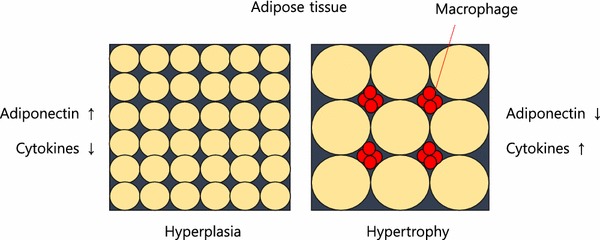
There are many causes of IR. Stress hormones (cortisol, adrenaline), inflammation, and high insulin levels themselves (response to dietary sugar and refined carbohydrates), each alone and in combination, cause immediate (within minutes to hours) insulin resistance. When these conditions persist over time insulin resistance becomes a chronic state. As fat cells grow in size, they reach a point where there is inadequate blood flow to the cells themselves and macrophages (immune cells that reside between the fat cells, most prominently in visceral fat) produce inflammatory chemicals called cytokines. Cytokines flow through the blood stream and effect every organ and every cell in the body creating a state of chronic inflammation which further worsens IR, creating a vicious cycle. As IR continues the pancreas produces increasingly higher amounts of insulin to maintain normal blood sugar levels but eventually IR becomes so great that blood sugar levels move into the “pre-diabetes” and eventually the diabetes range. IR builds for years to decades before blood sugar regulation fails. By the time blood sugar levels are “abnormal” insulin resistance has done great damage throughout the body.

Most doctors tragically do not order fasting insulin levels as routine blood tests. Fasting insulin levels rise long before fasting blood sugars and hemoglobin A1c start to rise. Meanwhile the damage progresses under the radar of routine testing.


1. Metabolic Engines: Muscle and Liver

Muscle

  • Normal Action: Insulin acts as a key that opens “doors” (GLUT4 receptors) to let glucose in for fuel. it also stimulates protein synthesis. Protein synthesis is essential to maintaining and increasing muscle mass and strength.
  • Insulin Resistance Effect: The “doors” stay locked. Glucose stays in the blood, and the muscle becomes “starched,” leading to sarcopenia (muscle wasting) and fatigue. The muscle can no longer utilize dietary protein to maintain or increase muscle mass.

Liver

  • Normal Action: Tells the liver to stop producing glucose and start storing it as glycogen or converting excess into fat.
  • Insulin Resistance Effect: The liver ignores the “stop” signal and keeps pumping out glucose while simultaneously ramping up fat production. This results in Non-Alcoholic Fatty Liver Disease (NAFLD).

2. Fat Cells (Adipose Tissue)

Visceral (Deep Fat) vs. Subcutaneous (Under Skin)

  • Normal Action: Insulin promotes fat storage and inhibits the breakdown of stored fat (lipolysis).
  • Insulin Resistance Effect: Fat cells—especially visceral ones—become “leaky.” They spill free fatty acids into the bloodstream and release inflammatory cytokines. This causes weight gain that is biologically difficult to lose because high insulin levels keep the “fat-burning” switch permanently off.

3. The Vital Organs: Heart, Kidneys, and Arteries

Heart and Arteries

  • Normal Action: Insulin stimulates the release of nitric oxide, which helps arteries relax and dilate.
  • Insulin Resistance Effect: Nitric oxide production drops, causing arteries to stiffen (hypertension). High insulin also damages the endothelial lining, leading to atherosclerosis (plaque buildup). This is the primary driver of heart failure, heart attacks and strokes.

Kidneys

  • Normal Action: Helps regulate sodium reabsorption.
  • Insulin Resistance Effect: The kidneys hold onto too much salt, increasing blood pressure. Over time, high blood sugar and inflammation damage the filtering units, leading to chronic kidney disease (CKD).

4. The Brain, Memory, and Sleep

Brain and Memory

  • Normal Action: Insulin crosses the blood-brain barrier to regulate appetite and support synaptic plasticity (the basis of learning).
  • Insulin Resistance Effect: Often called “Type 3 Diabetes,” brain IR starves neurons of energy and allows amyloid plaques and neurofibrillary tangles to build up. Worse, the brain is unable to utilize glucose to meet energy demands it starts to malfunction. This is a direct pathway to Alzheimer’s disease and dementia. As the small arteries in the brain become atherosclerotic and unable to deliver adequate oxygen and nourishment to brain cells small areas of the brain become permanently damaged eventually leading to vascular dementia.

Sleep

  • Insulin Resistance Effect: IR is heavily linked to Obstructive Sleep Apnea. (OSA) High insulin affects the central respiratory drive and increases fat deposits around the neck (a major contributor to obstructive sleep apnea), disrupting sleep cycles and creating periods of inadequate oxygen flow to the brain resulting in the acute stress response and awakening with each apneic event. Even without OSA, high insulin levels impair the production of melatonin which is essential to normal-restorative sleep. Throughout the day the brain accumulates metabolic toxins that must be cleared through the glymphatic system at night during sleep. As sleep is impaired this clearance system is disrupted, contributing to structural damage and functional loss. Sleep disruption and apneic episodes are stressful events, increasing stress hormones which then worsen IR, creating another vicious cycle. One night of sleep disruption causes acute IR. Chronic sleep disruption contributes to chronic IR.

5. Immunity and Structural Health

Immune System

  • Action: High insulin/glucose impairs white blood cell function.
  • Effect: Chronic inflammation (high CRP levels) and a weakened defense against infections. This is why diabetics often have poor wound healing. As normal immune regulation is impaired the immune system both over-reacts and under-reacts. Under-reaction increases risk of infection. Over-reaction produces cytokine storms seen with Covid-19 and other infections. Chronic inflammation worsens IR creating another vicious cycle. Chronic inflammation contributes to most chronic diseases.

Bone and Joints

  • Action: Insulin is bone-building.
  • Effect: IR leads to poor bone quality (despite high density) and osteoarthritis due to systemic inflammation and the “glycosylation” (sugar-coating) of joint cartilage, making it brittle.

6. The Pancreas: Beta and Alpha Cells

  • Normal Action: Beta cells produce insulin; Alpha cells produce glucagon (which raises sugar). They balance each other.
  • Insulin Resistance Effect:
    • Beta Cells: Work overtime to produce massive amounts of insulin to compensate, eventually “burning out” and dying. This can produce per4manent irreversible damage to the pancreas.
    • Alpha Cells: Become resistant to insulin’s “stop” signal and keep secreting glucagon, further raising blood sugar levels which in turn cause higher insulin secretion, both of which worsen IR, creating another vicious cycle.

7. Reproductive Effects: Infertility

  • In Women: High insulin stimulates the ovaries to produce excess testosterone, which is the primary driver of Polycystic Ovary Syndrome (PCOS) and infertility.
  • In Men: IR is a leading cause of low testosterone and erectile dysfunction (due to the arterial damage mentioned above).

Summary of Systemic Effects

ConditionPrimary Mechanism of Insulin Resistance
AtherosclerosisEndothelial dysfunction, high triglycerides, low HDL, increased TG/HDL ratio, increased small dense LDL and remnant particles, increased endothelial permeability.
DementiaNeuronal glucose starvation and plaque buildup, brain small vessel disease, disruption of blood brain barrier.
Chronic InflammationRelease of cytokines from visceral fat.
Heart FailureStiffening of the heart muscle and high blood pressure.
DiabetesPancreatic beta cell and alpha cell damage

Insulin’s Role vs. Insulin Resistance (IR)

Organ/SystemNormal Insulin ActionEffects of Insulin Resistance
LiverStops glucose production; stores glucose as glycogen.The liver ignores the “stop” signal, pumping out sugar even when you haven’t eaten (fatty liver).Fatty liver disease is the greatest cause of liver failure in the US.
MusclePrimary site for glucose uptake; promotes protein synthesis.Muscles can’t take in fuel efficiently, leading to fatigue and muscle wasting (sarcopenia). Muscle cells cannot use amino acids from dietary protein to maintain or build muscle. Elderly lose muscle and strength, resulting in falls, fractures and head trauma. Loss of muscle (the major sink for blood sugar after a meal) further increases duration and degree of blood sugar and insulin rise after a meal, which in turn increases IR. (vicious cycle)
Fat (Adipose)Stores fat; inhibits the breakdown of stored fat.Fat cells leak fatty acids into the blood, leading to high triglycerides and visceral fat gain. Macrophages (immune cells) produce inflammatory cytokines which circulate through the body contributing to chronic inflammation which worsens IR, another vicious cycle.
BrainRegulates appetite, memory, and cognitive function.Linked to “Type 3 Diabetes”; impaired memory and increased risk of neurodegeneration. Brain loses ability to meet energy demands and clear toxins. Insulin resistance in the brain explains memory loss, cognitive impairment, loss of neurons and synapses, loss of neuroplasticity. BDNF (brain derived neurotrophic factor) production is decreased by IR.
ArteriesStimulates nitric oxide for vasodilation (keeps vessels flexible).Reduced nitric oxide causes vessels to stiffen, raising blood pressure and plaque buildup. This is called endothelial dysfunction, the precursor to heart attack, stroke, peripheral vascular disease and a root cause for neuropathy and amputations.
HeartRegulates fuel use (switching between glucose and fats).The heart becomes “metabolically inflexible,” increasing the risk of heart failure.
KidneyManages sodium reabsorption and filtration.High insulin causes the kidneys to hold onto salt, driving up blood pressure and damaging filters. Oxidative stress leads to kidney failure.
Immune SystemModulates inflammation and helps T-cell function.Creates a state of “chronic low-grade inflammation” and weakens the response to infections.
BoneStimulates bone-forming cells (osteoblasts).Bone quality decreases; despite higher bone density in some cases, the bones are more brittle.
JointsMaintains cartilage and reduces systemic inflammation.High insulin promotes pro-inflammatory cytokines, accelerating osteoarthritis and gout.

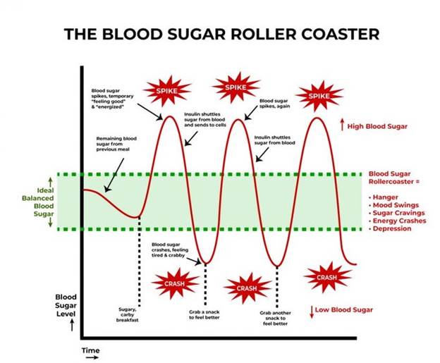
 A meal with sugar and refined carbohydrates causes excessive swings in blood sugar and insulin levels, creating insulin resistance and downstream damage. Alcohol consumption contributes to this process. Fat consumption does not cause a rise in blood sugar or insulin levels. Protein consumption produces a minimal rise in insulin levels in the absence of IR.

Fat storage can occur through hyperplasia (increase in number of fat cells) or hypertrophy (increase in size). Some ethnic groups are more prone to hypertrophy (south and east Asian). Hypertrophy in visceral fat (fat around the internal organs as opposed to fat under the skin) results in macrophage production of inflammatory cytokines. Eventually, the fat cells themselves can literally burst from too much volume.

 In my next post, I will discuss what we can do to prevent and reverse IR.

REFERENCES

Chadt A, Al-Hasani H. Glucose transporters in adipose tissue, liver, and skeletal muscle in metabolic health and disease. Pflugers Arch. 2020 Sep;472(9):1273-1298. doi: 10.1007/s00424-020-02417-x. Epub 2020 Jun 26. PMID: 32591906; PMCID: PMC7462924.

https://pmc.ncbi.nlm.nih.gov/articles/PMC7462924/

Fujita S, Rasmussen BB, Cadenas JG, Grady JJ, Volpi E. Effect of insulin on human skeletal muscle protein synthesis is modulated by insulin-induced changes in muscle blood flow and amino acid availability. Am J Physiol Endocrinol Metab. 2006 Oct;291(4):E745-54. doi: 10.1152/ajpendo.00271.2005. Epub 2006 May 16. PMID: 16705054; PMCID: PMC2804964.

https://pmc.ncbi.nlm.nih.gov/articles/PMC2804964

Vargas E, Joy NV, Carrillo Sepulveda MA. Biochemistry, Insulin Metabolic Effects. [Updated 2022 Sep 26]. In: StatPearls [Internet]. Treasure Island (FL): StatPearls Publishing; 2025 Jan-. Available from: https://www.ncbi.nlm.nih.gov/books/NBK525983/

https://www.ncbi.nlm.nih.gov/books/NBK525983/

Bugianesi E, Moscatiello S, Ciaravella MF, Marchesini G. Insulin resistance in nonalcoholic fatty liver disease. Curr Pharm Des. 2010 Jun;16(17):1941-51. doi: 10.2174/138161210791208875. PMID: 20370677.

https://pubmed.ncbi.nlm.nih.gov/20370677/

Cardillo C, Nambi SS, Kilcoyne CM, Choucair WK, Katz A, Quon MJ, Panza JA. Insulin stimulates both endothelin and nitric oxide activity in the human forearm. Circulation. 1999 Aug 24;100(8):820-5. doi: 10.1161/01.cir.100.8.820. PMID: 10458717.

https://pubmed.ncbi.nlm.nih.gov/10458717/

Ke JF, Wang JW, Zhang ZH, Chen MY, Lu JX, Li LX. Insulin Therapy Is Associated With an Increased Risk of Carotid Plaque in Type 2 Diabetes: A Real-World Study. Front Cardiovasc Med. 2021 Feb 1;8:599545. doi: 10.3389/fcvm.2021.599545. PMID: 33598483; PMCID: PMC7882504.

https://pubmed.ncbi.nlm.nih.gov/33598483/

Brosolo G, Da Porto A, Bulfone L, Vacca A, Bertin N, Scandolin L, Catena C, Sechi LA. Insulin Resistance and High Blood Pressure: Mechanistic Insight on the Role of the Kidney. Biomedicines. 2022 Sep 23;10(10):2374. doi: 10.3390/biomedicines10102374. PMID: 36289636; PMCID: PMC9598512.

https://pubmed.ncbi.nlm.nih.gov/36289636/

Kumar M, Dev S, Khalid MU, Siddenthi SM, Noman M, John C, Akubuiro C, Haider A, Rani R, Kashif M, Varrassi G, Khatri M, Kumar S, Mohamad T. The Bidirectional Link Between Diabetes and Kidney Disease: Mechanisms and Management. Cureus. 2023 Sep 20;15(9):e45615. doi: 10.7759/cureus.45615. PMID: 37868469; PMCID: PMC10588295.

https://pmc.ncbi.nlm.nih.gov/articles/PMC10588295/

Banks WA, Owen JB, Erickson MA. Insulin in the brain: there and back again. Pharmacol Ther. 2012 Oct;136(1):82-93. doi: 10.1016/j.pharmthera.2012.07.006. Epub 2012 Jul 17. PMID: 22820012; PMCID: PMC4134675.

https://pubmed.ncbi.nlm.nih.gov/22820012/

Rahman MS, Hossain KS, Das S, Kundu S, Adegoke EO, Rahman MA, Hannan MA, Uddin MJ, Pang MG. Role of Insulin in Health and Disease: An Update. Int J Mol Sci. 2021 Jun 15;22(12):6403. doi: 10.3390/ijms22126403. PMID: 34203830; PMCID: PMC8232639.

https://pmc.ncbi.nlm.nih.gov/articles/PMC8232639/

Scherrer U, Sartori C. Insulin as a vascular and sympathoexcitatory hormone: implications for blood pressure regulation, insulin sensitivity, and cardiovascular morbidity. Circulation. 1997 Dec 2;96(11):4104-13. doi: 10.1161/01.cir.96.11.4104. PMID: 9403636.

https://pubmed.ncbi.nlm.nih.gov/9403636/

Affuso F, Micillo F, Fazio S. Insulin Resistance, a Risk Factor for Alzheimer’s Disease: Pathological Mechanisms and a New Proposal for a Preventive Therapeutic Approach. Biomedicines. 2024 Aug 19;12(8):1888. doi: 10.3390/biomedicines12081888. PMID: 39200352; PMCID: PMC11351221.

https://pmc.ncbi.nlm.nih.gov/articles/PMC11351221/

Park MH, Kim DH, Lee EK, Kim ND, Im DS, Lee J, Yu BP, Chung HY. Age-related inflammation and insulin resistance: a review of their intricate interdependency. Arch Pharm Res. 2014 Dec;37(12):1507-14. doi: 10.1007/s12272-014-0474-6. Epub 2014 Sep 20. PMID: 25239110; PMCID: PMC4246128.

https://pubmed.ncbi.nlm.nih.gov/25239110/

Hardy OT, Czech MP, Corvera S. What causes the insulin resistance underlying obesity? Curr Opin Endocrinol Diabetes Obes. 2012 Apr;19(2):81-7. doi: 10.1097/MED.0b013e3283514e13. PMID: 22327367; PMCID: PMC4038351.

https://pmc.ncbi.nlm.nih.gov/articles/PMC4038351/

THIS WEBSITE PROVIDES INFORMATION FOR EDUCATIONAL PURPOSES ONLY. CONSULT YOUR HEALTH CARE PROVIDER FOR MEDICAL ADVICE.

Eat clean, drink filtered water, love, laugh, exercise outdoors in a greenspace, get some morning sunlight, block the blue light before bed, engage in meaningful work, find a sense of purpose, spend time with those you love, AND sleep well tonight.

Doctor Bob

Normal Thyroid function tests (T4 and TSH) do not guarantee you have normal thyroid function.

In his book Blindspots, Marty Makary MD (slated to be the next FDA commissioner) discusses why routine medical practices are often slow to change, despite mounting evidenced that those practices are not supported by best evidence. Although not discussed in his book, the evaluation and treatment of hypothyroidism presents a prime example of this problem. Before diving into the issues, lets review some basic physiology.

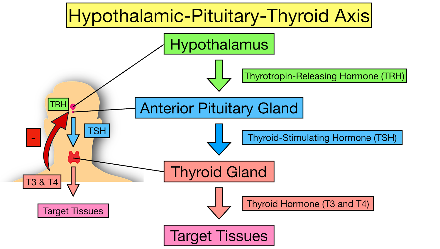
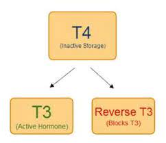
The hypothalamus in the brain produces TRH (thyrotropin releasing hormone) in response to circulating levels of T3 and T4 (two forms of thyroid hormone). TRH then stimulates production and release of TSH (thyroid stimulating hormone) in the pituitary gland. In turn, TSH stimulates the thyroid gland to produce thyroid hormone. The two forms of thyroid hormone produced by the thyroid gland are called T4 and T3 (T3 has 3 iodine molecules, T4 has 4) T4 is the predominant hormone produced by the thyroid gland. In the liver and the kidney T4 is converted to T3 through a process called deiodination, supplying 80% of circulating T3. Importantly, T3 is 7 to 10 times more potent than T4.

Both T3 and T4 directly inhibit TSH synthesis. This feedback system helps maintain homeostasis under conditions of optimal health. (https://www.ncbi.nlm.nih.gov/books/NBK278958/)

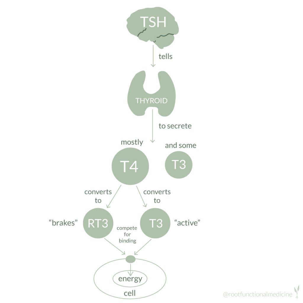
Taken from: https://rootfunctionalmedicine.com/conversion-of-t4-to-t3-thyroid-hormone/

When testing thyroid function, most physicians order only TSH (thyroid stimulating hormone) and T4. But to have normal thyroid function several conditions must be met. Of those many conditions, three are often not considered by most physicians and not evaluated with TSH and T4. First, there must be adequate conversion of T4 into T3, because T3 is the form of thyroid hormone that provides most physiologic effect. Second, there must be adequate transport of T4 and T3 into your cells. Third, levels of reverse T3 should not be high enough to block adequate amounts of Thyroid Hormone binding to receptors on your cells.

The “normal” range of TSH in most labs is 0.4 to 5.0 mU/L. Recently a narrower range of 0.5-2.5 mU/L has been proposed to exclude individuals with “minimal” thyroid dysfunction, but this is controversial. This controversy is the crux of one of the many problems in interpreting “normal” thyroid function tests. Proponents of the narrower range maintain that patients with symptoms of hypothyroidism and a TSH above 2.5 might benefit from treatment. In addition, proponents of the narrower range suggest that some patients with TSH levels above 2.5 may not report symptoms of hypothyroidism but their physiology may be suboptimal for health. (https://www.ncbi.nlm.nih.gov/books/NBK278958/) Physiology of the Hypothalamic-Pituitary-Thyroid Axis.

Treatment with T4 alone, may not provide adequate thyroid replacement.

If a physician orders only T4 and TSH to evaluate thyroid function, but conversion of T4 to T3 is impaired, a normal T4 and normal TSH will be interpreted as normal thyroid function. But with low T3 levels, a patient can experience symptoms of hypothyroidism. Likewise, if a patient is only treated with T4 (levothyroxine) but has issues converting T4 to T3, they may need to receive a prescription for T3 and T4. Using the ratio of Free T3 to Free T4 one can identify patients with problems caused by inadequate deiodination (conversion of T4 to T3). Monotherapy with levothyroxine (T4) has been standard of care for decades. But studies have demonstrated that patients treated with T4 only have relatively low serum levels of T3 (tri-iodothyronine) compared to the general population and symptoms of hypothyroidism persist for some patients despite normal TSH levels.

Issues of active versus passive transport.

T3 and T4 enter the hypothalamus and pituitary glands “passively”, meaning an active transport mechanism is not necessary. But in the rest of the body active transport of T3 and T4 into cells is necessary. If there is a problem with the active transport system, the hypothalamus and pituitary may “see” normal levels of T3 and T4 but the rest of the body may not be getting the full benefit of thyroid hormone. Thus, TSH levels will be normal, T4 levels will be normal, but cellular T3 and T4 levels will not be adequate. Yet physicians will interpret a normal TSH and T4 to mean normal thyroid function. Conditions that impair thyroid hormone transport into cells include: insulin resistance, diabetes, obesity, chronic and acute dieting, diabetes, depression, anxiety, bipolar disorder, neurodegenerative diseases, chronic fatigue syndrome, fibromyalgia, cardiovascular disease, inflammation and chronic illness, and disorders of lipid metabolism.

Reverse T3 can cause problems.

High levels of reverse T3 can occur under many conditions including chronic stress, chronic inflammation and many chronic diseases. As previously discussed, Reverse T3 binds to Thyroid hormone receptors but does not have the beneficial effect of normal T3, thus blocking T3 from doing its job. Most physicians do not measure reverse T3 or calculate the T3/reverse T3 ratio.

Therefore, in addition to problems with deiodinase (conversion of T4 to T3), high levels of reverse t3 can render monotherapy with levothyroxine inadequate, while leaving TSH and T4 levels “normal”.

How to calculate t3/reverse t3 ratio – National Academy of Hypothyroidism

This has been a quick discussion of a complex system. Not all aspects of thyroid testing and treatment have been addressed. Hopefully this discussion will help you understand why simply measuring TSH and T4 (a common practice) will not tell you whether you have normal thyroid function. Ideally, all patients being tested would have free T3 and reverse T3 measured in addition to T4 and TSH. In addition, the free T3/reverse T3 ratio and the free T3/freeT4 ratio would be considered when deciding whether treatment with T4 alone (levothyroxine monotherapy) is adequate. Many patients would benefit from adding T3 (triiodothyronine) to T4 (levothyroxine) therapy. In addition, a TSH level above 2.5 should be carefully evaluated for hypothyroid symptoms as levels above 2.5 are arguably “abnormal” (or at least should raise a red flag as to the possibility). Finally, recognize that this discussion presents controversies in medicine. Most practitioners apply the concept of “sick euthyroid” to patients with acute illness and low T3 levels, considering it a “normal” protective mechanism that does not require thyroid hormone therapy. But under chronic conditions, many practitioners who think “outside the box” would consider poor conversion of T4 to T3 and/or high levels of reverse T3, as possible indications for thyroid hormone therapy when symptoms of hypothyroidism are present. Under these circumstances directly addressing underlying causes such as chronic inflammation may provide the best initial approach. (anti-inflammatory diet, regular exercise, good sleep habits, stress reduction, strengthen social support)

But when a patient is already on monotherapy with T4, consideration of adding T3 to improve quality of life and physiologic function is worthy of consideration when comprehensive laboratory evaluation suggests problems as described above (cellular transport, T4 to T3 conversion, high reverse T3).

For a greater dive into this subject follow these links.

Physiology of the Hypothalamic-Pituitary-Thyroid Axis.

The relevance of T3 in the management of hypothyroidism – The Lancet Diabetes & Endocrinology

Levothyroxine Monotherapy Cannot Guarantee Euthyroidism in All Athyreotic Patients

Is a Normal TSH Synonymous With “Euthyroidism” in Levothyroxine Monotherapy?

Thyroid Hormone Transport into cellular tissue.

HPA axis dysfunction in Chronic Fatigue Syndrome and Fibromyalgia

Hormone replacement therapy in the geriatric patient, part 1.

Hormone replacement therapy in the geriatric patient, part 2.

Peripheral thyroid hormone conversion.

What is reverse T3 syndrome?

A complete pathway map of T4 and T3 metabolism and clearance

The Low T3 syndrome

THIS WEBSITE PROVIDES INFORMATION FOR EDUCATIONAL PURPOSES ONLY. CONSULT YOUR HEALTH CARE PROVIDER FOR MEDICAL ADVICE.

Eat clean, drink filtered water, love, laugh, exercise outdoors in a greenspace, get some morning sunlight, block the blue light before bed, engage in meaningful work, find a sense of purpose, spend time with those you love, AND sleep well tonight.

Doctor Bob