Category Archives: alzheimer’s disease

Can a drug replace the benefits of exercise?

A new drug, still under investigation and not yet FDA approved is reported to provide the benefits of exercise in humans. The drug is called ATX304 and has been studied in humans in phase II clinical trials. It works by decreasing the degradation of a master energy controller called AMPK (AMP-activated protein kinase) By decreasing the degradation, it increases the amount of AMPK which has multiple favorable metabolic effects. When we exercise our muscles utilize ATP (adenosine triphosphate) the major energy source for our cells. When ATP is broken down the levels of AMP (adenosine monophosphate) increase and that increase signals AMPK to increase. ATX304 produces the same rise in AMPK experienced with exercise. The result is that muscle cells uptake glucose from the circulation without the need for insulin (non insulin dependent glucose uptake), the same occurs with exercise. With exercise the effect can last for 16 hours. Thus blood sugar is better controlled with both exercise and with ATX304. Along with better blood sugar control, insulin sensitivity is maintained and improved by avoiding pancreatic beta cell stress (beta cells produce insulin). ATX-304 also improves blood vessel flow, cardiac pump function, and as a result improved delivery of oxygen and nutrients to muscle and all parts of the body.

ATX304 also increases the uptake of fatty acids by the mitochondria (the organelle in our cells that produce energy in the form of ATP), facilitating energy production through the burning of fat (a process called beta oxidation). This happens without the muscle loss associated with GLP-1 drugs. Muscle is protected because glucose and fatty acid delivery to muscles are enhanced while keeping cellular ATP stable, completely bypassing the starvation pathways that cause muscle wasting when people lose weight by fasting or taking GLP-1 agonist drugs.

While I do not advocate taking pills instead of exercise I view ATX-304 as a better alternative weight loss medication compared to GLP-1 drugs for several reasons. First and foremost one can avoid the 20-40% loss of muscle associated with GLP-1 drugs. Secondly, ATX-304 increases metabolic rate, causing greater energy expenditure.

While ATX-304 represents an exciting potential approach to weight loss while improving insulin sensitivity without loss of muscle, you cannot build muscle strength, muscle mass, improve balance and coordination, or challenge your brain (with the resulting cognitive protection) by taking a pill. You must exercise including resistance training to accomplish those specific beneficial outcomes.

For a deeper dive into ATX-304 and a more detailed understanding how our bodies use glucose and fat to produce energy, as well as understanding how exercise works to improve metabolic health, what follows is a conversation I had with Google Gemini.

Conversation with Gemini

how does atx304 improve glucose control, enhance exercise performance and reduce body fat while maintaining muscle, use only peer reviewed scientific papers and cite references

ATX-304 (formerly known as O304) is a first-in-class, small-molecule, non-allosteric PAN-AMPK activator (AMP-activated protein kinase) currently in clinical development for metabolic and cardiovascular diseases (Ericsson et al., 2021; Steneberg et al., 2018). It functions primarily by protecting phosphorylated AMPK (p-AMPK) from dephosphorylation by protein phosphatases like PP2C, effectively acting as an “exercise mimetic” that triggers catabolic, energy-generating pathways (Steneberg et al., 2018).

Peer-reviewed academic research details the distinct physiological mechanisms through which ATX-304 improves glucose control, enhances exercise performance, and selectively targets body fat while preserving lean muscle mass:

1. Improvement of Glucose Control

ATX-304 manages blood sugar levels through a dual mechanism targeting both skeletal muscle and pancreatic tissues:

  • Insulin-Independent Glucose Uptake: In skeletal muscle, ATX-304 activates AMPK signaling, which drives the translocation of glucose transporter 4 (GLUT4) vesicles to the cell membrane. This allows muscle tissue to pull glucose from the bloodstream independently of insulin action (Steneberg et al., 2018).
  • Beta-Cell Preservation and Rest: By reducing systemic glucose demand and chronic hyperglycemia, the compound mitigates pancreatic β-cell stress. Clinical evidence from Phase IIa trials in patients with Type 2 Diabetes (T2D) demonstrated substantial reductions in fasting plasma glucose (FPG) and overall insulin resistance, quantified by HOMA-IR (Steneberg et al., 2018).
  • Reversal of Age-Related Insulin Resistance: In longitudinal animal models, prolonged administration of the compound prevented the progressive, age-associated hyperinsulinemia that typically induces metabolic syndrome (Ericsson et al., 2021).

2. Enhancement of Exercise Performance

As an exercise mimetic, ATX-304 induces cellular adaptations similar to endurance training:

  • Cardiovascular & Microvascular Remodeling: ATX-304 mimics the cardiac stressors of exercise by enhancing left ventricular stroke volume, increasing end-diastolic volume, and lowering resting heart rate without inducing pathological cardiac hypertrophy (Ericsson et al., 2021; Steneberg et al., 2018). Furthermore, it improves peripheral microvascular perfusion and blood pressure regulations.
  • Mitochondrial Homeostasis: The compound enhances mitochondrial respiration, upregulates fatty acid oxidation, and increases baseline oxygen consumption rates (OCR) in metabolic tissues (Katerelos et al., 2024). These changes directly translate to significantly improved exercise capacity and total work endurance, especially in aged or metabolically compromised subjects (Ericsson et al., 2021).

3. Reduction of Body Fat While Maintaining Muscle

One of the key advantages of direct pan-AMPK activation via ATX-304 is its ability to execute a systemic “metabolic switch” that differentiates between fat tissue and lean body mass:

  • Upregulation of Lipid Oxidation: ATX-304 stimulates cellular catabolism by phosphorylating Acetyl-CoA Carboxylase (ACC). Phosphorylation inhibits ACC, which decreases malonyl-CoA levels, disinhibits carnitine palmitoyltransferase-1 (CPT1), and permits rapid fatty acid oxidation in both the liver and adipose tissue (Holm et al., 2024).
  • Selective Adiposity Loss: Body composition analysis via EchoMRI reveals that the rapid weight reduction induced by ATX-304 corresponds almost entirely to a loss in adipose (fat) mass (Holm et al., 2024). It also limits hepatic de novo lipogenesis and drives down blood cholesterol levels.
  • Muscle Preservation (Lean Weight Maintenance): Unlike severe caloric restriction or certain anti-obesity medications that trigger concurrent muscle loss, animals treated with ATX-304 maintain their absolute lean weight (muscle mass) (Holm et al., 2024). The compound drives a significant long-term increase in energy expenditure, meaning that even when food intake naturally rebounds or increases by 20% to 30%, fat mass continues to decrease while skeletal muscle architecture is protected (Holm et al., 2024).

References

Ericsson, M., Steneberg, P., Nyrén, R., & Edlund, H. (2021). AMPK activator O304 improves metabolic and cardiac function, and exercise capacity in aged mice. Communications Biology, 4(1), 1–12. https://doi.org/10.1038/s42003-021-02837-0 Cited by: 30

Holm, E., Vermeulen, I., Parween, S., López-Pérez, A., Cillero-Pastor, B., Vandenbosch, M., Remeseiro, S., & Hörnblad, A. (2024). AMPK-activator ATX-304 reduces oxidative stress and improves MASLD via metabolic switching. JCI Insight, 9(7). https://doi.org/10.1101/2024.02.13.578901 Cited by: 10

Katerelos, M., Gleich, K., Harley, G., Loh, K., Oakhill, J. S., Kemp, B. E., de Souza, D. P., Narayana, V. K., Coughlan, M. T., Laskowski, A., Ling, N. X. Y., Murray-Segal, L., Brink, R., Lee, M., Power, D. A., & Mount, P. F. (2024). The AMPK activator ATX-304 alters cellular metabolism to protect against cisplatin-induced acute kidney injury. Biomedicine & Pharmacotherapy, 175, 116730. https://doi.org/10.1016/j.biopha.2024.116730 Cited by: 9

Steneberg, P., Lindahl, E., Dahl, U., Lidh, E., Straseviciene, J., Backlund, F., Kjellkvist, E., Berggren, E., Lundberg, I., Bergqvist, I., Ericsson, M., Eriksson, B., Linde, K., Westman, J., Edlund, T., & Edlund, H. (2018). PAN-AMPK activator O304 improves glucose homeostasis and microvascular perfusion in mice and type 2 diabetes patients. JCI Insight, 3(12). https://doi.org/10.1172/jci.insight.99114 Cited by: 132

how do the effects on ACC and CPT1 lead to increased fat oxidation

The biochemical mechanism linking Acetyl-CoA Carboxylase (ACC) and Carnitine Palmitoyltransferase-1 (CPT1) is the primary pathway through which AMPK activators like ATX-304 accelerate fat oxidation.

In cellular metabolism, CPT1 is the “gatekeeper” for fat burning, but its activity is strictly regulated by a molecular brake called malonyl-CoA. Direct activation of AMPK breaks this cycle, shifting the cell into a dedicated fat-burning state.

The Molecular Chain Reaction

When ATX-304 activates AMPK, it initiates a sequential cascade that directly impacts the mitochondria:

        ATX-304 / AMPK Activated

                   │

                   ▼

         ACC is Phosphorylated

             (Inactivated)

                   │

                   ▼

       Malonyl-CoA Levels Drop

         (The “Brake” is Removed)

                   │

                   ▼

        CPT1 is Disinhibited

             (Gate Opens)

                   │

                   ▼

 Fatty Acids Enter Mitochondria ──► Beta-Oxidation (Fat Burning)

1. The Inactivation of ACC

Under normal conditions, the enzyme ACC converts acetyl-CoA into malonyl-CoA. Malonyl-CoA is the fundamental building block for synthesizing new fatty acids (de novo lipogenesis).

When ATX-304 activates AMPK, AMPK attaches a phosphate group to ACC (specifically the ACC1 and ACC2 isoforms). This phosphorylation inactivates ACC, halting the production of malonyl-CoA.

2. The Disinhibition of CPT1

Malonyl-CoA acts as a powerful allosteric inhibitor (a biological “off switch”) for CPT1. CPT1 is an enzyme anchored to the outer mitochondrial membrane. Long-chain fatty acids cannot cross into the mitochondria on their own; they must be attached to carnitine by CPT1 to gain entry.

Because ATX-304 shuts down ACC, cellular levels of malonyl-CoA rapidly drop. Without malonyl-CoA around to inhibit it, CPT1 transitions into its fully active state.

3. Increased Mitochondrial Beta-Oxidation

With the CPT1 gateway open, long-chain fatty acids are rapidly converted into acyl-carnitines, allowing them to pass through the mitochondrial membranes via the carnitine shuttle. Once inside the mitochondrial matrix, these fatty acids undergo β-oxidation (beta-oxidation).

During β-oxidation, the fatty acids are chopped down into acetyl-CoA molecules, which directly feed into the Tricarboxylic Acid (TCA) cycle to generate ATP (cellular energy).

Summary: By turning off ACC, ATX-304 depletes the molecule (malonyl-CoA) that normally blocks CPT1. This unlocks the mitochondrial gateway, forcing the cell to pull circulating fatty acids out of storage and burn them for fuel, ultimately driving down systemic body fat (Holm et al., 2024; Steneberg et al., 2018).

Want to know how this specific fat-burning pathway preserves muscle mass?

Yes

How does ATX-304 increase fat oxidation via ACC and CPT1 without triggering skeletal muscle breakdown or protein degradation?

The preservation of skeletal muscle mass during accelerated fat oxidation is a defining feature of ATX-304. Typically, extreme caloric restriction or non-selective metabolic stressors that force fat burning also trigger a counter-regulatory catabolic state, where muscle tissue is broken down for amino acids.

ATX-304 avoids skeletal muscle breakdown through several distinct molecular and metabolic safeguards:

1. Preservation of Cellular ATP Levels

diabetesjournals.org

Traditional methods of AMPK activation (like mitochondrial poisons or extreme energy deprivation) severely deplete cellular ATP. When ATP drops drastically, hyper-activated AMPK signals the cell that it is starving, which triggers the ubiquitin-proteasome system (via MuRF1 and MAFbx/Atrogin-1) and FoxO3-mediated autophagy to degrade muscle protein for emergency energy.

  • The ATX-304 Difference: ATX-304 activates AMPK by preventing the dephosphorylation of p-AMPK by protein phosphatases, rather than by draining the cell of energy (Steneberg et al., 2018).

http://www.researchgate.net

  • Peer-reviewed data confirms that cellular ATP levels and mitochondrial membrane potential (MMP) are fully preserved under ATX-304 exposure (Diabetes, 2026). Because the muscle cell is never in a true energetic crisis or ATP deficit, the signals that initiate muscle proteolysis (protein degradation) are never switched on.

diabetesjournals.org

2. Enhanced Glucose and Nutrient Delivery to Muscle

insight.jci.org

Muscle wasting often occurs when a lack of localized energy forces muscle fibers to degrade. ATX-304 directly protects muscle tissue by optimizing its nutrient environment:

  • GLUT4 Translocation: In skeletal muscle, ATX-304-driven AMPK activation stimulates the moving of GLUT4 transporters to the cell membrane, enhancing insulin-independent glucose uptake (Steneberg et al., 2018).

http://www.baybridgebio.com

  • Microvascular Perfusion: The compound improves capillary blood flow and microvascular perfusion directly to skeletal muscle tissue. This ensures a continuous supply of blood sugar, oxygen, and systemic nutrients to the muscle, keeping the tissue fueled and preventing the need for autophagic self-cannibalization.

3. Metabolic Substrate Switching Away from Proteolysis

By targeting the ACC-CPT1 axis, ATX-304 forces a highly selective “metabolic switch.”

  • The inhibition of ACC stops fatty acid synthesis and drops malonyl-CoA levels, allowing CPT1 to constantly shuttle long-chain fatty acids into the mitochondria (Holm et al., 2024).
  • This floods the mitochondrial matrix with lipids for β-oxidation. Because the cell is over-supplied with lipid-derived acetyl-CoA to feed the TCA cycle, it heavily relies on fat as its primary fuel source. Consequently, there is no biochemical demand to oxidize branched-chain amino acids (BCAAs) from structural muscle proteins.

4. Absence of the “Starvation Deficit” Loop

In long-term animal studies, subjects treated with ATX-304 experienced a substantial reduction in fat mass while maintaining absolute lean body weight (Holm et al., 2024). Because ATX-304 increases baseline energy expenditure through increased mitochondrial respiration, treated subjects naturally maintain a normal or slightly increased dietary intake to compensate. This stands in sharp contrast to crash dieting or GLP-1 receptor agonists, where profound appetite suppression and extreme caloric deficits frequently result in up to 20–40% of the lost weight coming from lean muscle mass.

http://www.researchgate.net

In short: ATX-304 accelerates fat burning through the ACC-CPT1 pathway without sacrificing muscle because it increases lipid availability and muscle glucose uptake while keeping cellular ATP stable—completely bypassing the starvation pathways that cause muscle wasting.

References

  • Holm, E., et al. (2024). AMPK-activator ATX-304 reduces oxidative stress and improves MASLD via metabolic switching. JCI Insight, 9(7).

http://www.biorxiv.org

  • Steneberg, P., et al. (2018). PAN-AMPK activator O304 improves glucose homeostasis and microvascular perfusion in mice and type 2 diabetes patients. JCI Insight, 3(12).

pmc.ncbi.nlm.nih.gov

  • American Diabetes Association (ADA) Scientific Sessions. (2026). Abstract 1788-P: AMPK Activation by ATX-304 Is Not Secondary to Changes in ATP Levels. Diabetes, 75(Supplement 1).

diabetesjournals.org+ 1

THIS WEBSITE PROVIDES INFORMATION FOR EDUCATIONAL PURPOSES ONLY. CONSULT YOUR HEALTH CARE PROVIDER FOR MEDICAL ADVICE.

Eat clean, drink filtered water, love, laugh, exercise outdoors in a greenspace, get some morning sunlight, block the blue light before bed, engage in meaningful work, find a sense of purpose, spend time with those you love, AND sleep well tonight.

Doctor Bob
Insulin Resistance, the silent killer and root cause of modern chronic disease.

Insulin is much more than a blood sugar hormone. Produced by the pancreas primarily in response to carbohydrate and sugar consumption, insulin is a master anabolic signal that dictates how every cell in your body grows, uses energy, and repairs itself. When insulin levels are healthy, it keeps the body in a state of “build and store.” When insulin resistance (IR) develops, the body loses its ability to hear this signal, leading to systemic breakdown. Instead of “build and store” the body deteriorates, causing loss of muscle mass, strength, energy production, memory and cognitive function, bone strength, brain cells and connections, ability of blood vessels to relax, ability for the heart to pump blood, ability to achieve restorative sleep, ability of the liver and kidneys to clear toxins from the body, even the ability to reproduce resulting in infertility and erectile dysfunction. Visceral fat stores increase to destructive levels resulting in obesity and obesity-related complications including chronic inflammation which further drives IR to higher levels.

IR is a root cause of cardiovascular disease (heart attack, stroke, hypertension, heart failure), many kinds of cancer (directly linked to breast, prostate and colon cancer), kidney failure, heart failure, dementia, osteoporosis, osteoarthritis, and much more.  IR is causally linked or a contributor to, every chronic non-communicable disease of modern civilization.

WHAT IS INSULIN RESISTANCE?

Insulin resistance is the inability of cells and organs to respond normally to insulin signaling. Every cell of every organ has insulin receptors that initiate action by the cell and organ.

WHAT CAUSES INSULIN RESISTANCE?

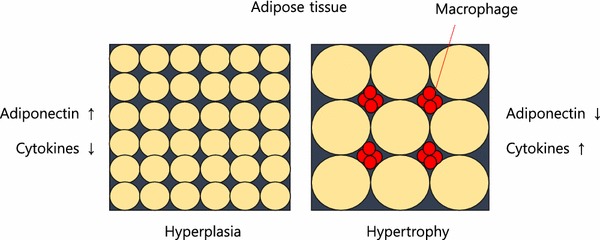
There are many causes of IR. Stress hormones (cortisol, adrenaline), inflammation, and high insulin levels themselves (response to dietary sugar and refined carbohydrates), each alone and in combination, cause immediate (within minutes to hours) insulin resistance. When these conditions persist over time insulin resistance becomes a chronic state. As fat cells grow in size, they reach a point where there is inadequate blood flow to the cells themselves and macrophages (immune cells that reside between the fat cells, most prominently in visceral fat) produce inflammatory chemicals called cytokines. Cytokines flow through the blood stream and effect every organ and every cell in the body creating a state of chronic inflammation which further worsens IR, creating a vicious cycle. As IR continues the pancreas produces increasingly higher amounts of insulin to maintain normal blood sugar levels but eventually IR becomes so great that blood sugar levels move into the “pre-diabetes” and eventually the diabetes range. IR builds for years to decades before blood sugar regulation fails. By the time blood sugar levels are “abnormal” insulin resistance has done great damage throughout the body.

Most doctors tragically do not order fasting insulin levels as routine blood tests. Fasting insulin levels rise long before fasting blood sugars and hemoglobin A1c start to rise. Meanwhile the damage progresses under the radar of routine testing.


1. Metabolic Engines: Muscle and Liver

Muscle

  • Normal Action: Insulin acts as a key that opens “doors” (GLUT4 receptors) to let glucose in for fuel. it also stimulates protein synthesis. Protein synthesis is essential to maintaining and increasing muscle mass and strength.
  • Insulin Resistance Effect: The “doors” stay locked. Glucose stays in the blood, and the muscle becomes “starched,” leading to sarcopenia (muscle wasting) and fatigue. The muscle can no longer utilize dietary protein to maintain or increase muscle mass.

Liver

  • Normal Action: Tells the liver to stop producing glucose and start storing it as glycogen or converting excess into fat.
  • Insulin Resistance Effect: The liver ignores the “stop” signal and keeps pumping out glucose while simultaneously ramping up fat production. This results in Non-Alcoholic Fatty Liver Disease (NAFLD).

2. Fat Cells (Adipose Tissue)

Visceral (Deep Fat) vs. Subcutaneous (Under Skin)

  • Normal Action: Insulin promotes fat storage and inhibits the breakdown of stored fat (lipolysis).
  • Insulin Resistance Effect: Fat cells—especially visceral ones—become “leaky.” They spill free fatty acids into the bloodstream and release inflammatory cytokines. This causes weight gain that is biologically difficult to lose because high insulin levels keep the “fat-burning” switch permanently off.

3. The Vital Organs: Heart, Kidneys, and Arteries

Heart and Arteries

  • Normal Action: Insulin stimulates the release of nitric oxide, which helps arteries relax and dilate.
  • Insulin Resistance Effect: Nitric oxide production drops, causing arteries to stiffen (hypertension). High insulin also damages the endothelial lining, leading to atherosclerosis (plaque buildup). This is the primary driver of heart failure, heart attacks and strokes.

Kidneys

  • Normal Action: Helps regulate sodium reabsorption.
  • Insulin Resistance Effect: The kidneys hold onto too much salt, increasing blood pressure. Over time, high blood sugar and inflammation damage the filtering units, leading to chronic kidney disease (CKD).

4. The Brain, Memory, and Sleep

Brain and Memory

  • Normal Action: Insulin crosses the blood-brain barrier to regulate appetite and support synaptic plasticity (the basis of learning).
  • Insulin Resistance Effect: Often called “Type 3 Diabetes,” brain IR starves neurons of energy and allows amyloid plaques and neurofibrillary tangles to build up. Worse, the brain is unable to utilize glucose to meet energy demands it starts to malfunction. This is a direct pathway to Alzheimer’s disease and dementia. As the small arteries in the brain become atherosclerotic and unable to deliver adequate oxygen and nourishment to brain cells small areas of the brain become permanently damaged eventually leading to vascular dementia.

Sleep

  • Insulin Resistance Effect: IR is heavily linked to Obstructive Sleep Apnea. (OSA) High insulin affects the central respiratory drive and increases fat deposits around the neck (a major contributor to obstructive sleep apnea), disrupting sleep cycles and creating periods of inadequate oxygen flow to the brain resulting in the acute stress response and awakening with each apneic event. Even without OSA, high insulin levels impair the production of melatonin which is essential to normal-restorative sleep. Throughout the day the brain accumulates metabolic toxins that must be cleared through the glymphatic system at night during sleep. As sleep is impaired this clearance system is disrupted, contributing to structural damage and functional loss. Sleep disruption and apneic episodes are stressful events, increasing stress hormones which then worsen IR, creating another vicious cycle. One night of sleep disruption causes acute IR. Chronic sleep disruption contributes to chronic IR.

5. Immunity and Structural Health

Immune System

  • Action: High insulin/glucose impairs white blood cell function.
  • Effect: Chronic inflammation (high CRP levels) and a weakened defense against infections. This is why diabetics often have poor wound healing. As normal immune regulation is impaired the immune system both over-reacts and under-reacts. Under-reaction increases risk of infection. Over-reaction produces cytokine storms seen with Covid-19 and other infections. Chronic inflammation worsens IR creating another vicious cycle. Chronic inflammation contributes to most chronic diseases.

Bone and Joints

  • Action: Insulin is bone-building.
  • Effect: IR leads to poor bone quality (despite high density) and osteoarthritis due to systemic inflammation and the “glycosylation” (sugar-coating) of joint cartilage, making it brittle.

6. The Pancreas: Beta and Alpha Cells

  • Normal Action: Beta cells produce insulin; Alpha cells produce glucagon (which raises sugar). They balance each other.
  • Insulin Resistance Effect:
    • Beta Cells: Work overtime to produce massive amounts of insulin to compensate, eventually “burning out” and dying. This can produce per4manent irreversible damage to the pancreas.
    • Alpha Cells: Become resistant to insulin’s “stop” signal and keep secreting glucagon, further raising blood sugar levels which in turn cause higher insulin secretion, both of which worsen IR, creating another vicious cycle.

7. Reproductive Effects: Infertility

  • In Women: High insulin stimulates the ovaries to produce excess testosterone, which is the primary driver of Polycystic Ovary Syndrome (PCOS) and infertility.
  • In Men: IR is a leading cause of low testosterone and erectile dysfunction (due to the arterial damage mentioned above).

Summary of Systemic Effects

ConditionPrimary Mechanism of Insulin Resistance
AtherosclerosisEndothelial dysfunction, high triglycerides, low HDL, increased TG/HDL ratio, increased small dense LDL and remnant particles, increased endothelial permeability.
DementiaNeuronal glucose starvation and plaque buildup, brain small vessel disease, disruption of blood brain barrier.
Chronic InflammationRelease of cytokines from visceral fat.
Heart FailureStiffening of the heart muscle and high blood pressure.
DiabetesPancreatic beta cell and alpha cell damage

Insulin’s Role vs. Insulin Resistance (IR)

Organ/SystemNormal Insulin ActionEffects of Insulin Resistance
LiverStops glucose production; stores glucose as glycogen.The liver ignores the “stop” signal, pumping out sugar even when you haven’t eaten (fatty liver).Fatty liver disease is the greatest cause of liver failure in the US.
MusclePrimary site for glucose uptake; promotes protein synthesis.Muscles can’t take in fuel efficiently, leading to fatigue and muscle wasting (sarcopenia). Muscle cells cannot use amino acids from dietary protein to maintain or build muscle. Elderly lose muscle and strength, resulting in falls, fractures and head trauma. Loss of muscle (the major sink for blood sugar after a meal) further increases duration and degree of blood sugar and insulin rise after a meal, which in turn increases IR. (vicious cycle)
Fat (Adipose)Stores fat; inhibits the breakdown of stored fat.Fat cells leak fatty acids into the blood, leading to high triglycerides and visceral fat gain. Macrophages (immune cells) produce inflammatory cytokines which circulate through the body contributing to chronic inflammation which worsens IR, another vicious cycle.
BrainRegulates appetite, memory, and cognitive function.Linked to “Type 3 Diabetes”; impaired memory and increased risk of neurodegeneration. Brain loses ability to meet energy demands and clear toxins. Insulin resistance in the brain explains memory loss, cognitive impairment, loss of neurons and synapses, loss of neuroplasticity. BDNF (brain derived neurotrophic factor) production is decreased by IR.
ArteriesStimulates nitric oxide for vasodilation (keeps vessels flexible).Reduced nitric oxide causes vessels to stiffen, raising blood pressure and plaque buildup. This is called endothelial dysfunction, the precursor to heart attack, stroke, peripheral vascular disease and a root cause for neuropathy and amputations.
HeartRegulates fuel use (switching between glucose and fats).The heart becomes “metabolically inflexible,” increasing the risk of heart failure.
KidneyManages sodium reabsorption and filtration.High insulin causes the kidneys to hold onto salt, driving up blood pressure and damaging filters. Oxidative stress leads to kidney failure.
Immune SystemModulates inflammation and helps T-cell function.Creates a state of “chronic low-grade inflammation” and weakens the response to infections.
BoneStimulates bone-forming cells (osteoblasts).Bone quality decreases; despite higher bone density in some cases, the bones are more brittle.
JointsMaintains cartilage and reduces systemic inflammation.High insulin promotes pro-inflammatory cytokines, accelerating osteoarthritis and gout.

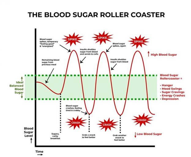
 A meal with sugar and refined carbohydrates causes excessive swings in blood sugar and insulin levels, creating insulin resistance and downstream damage. Alcohol consumption contributes to this process. Fat consumption does not cause a rise in blood sugar or insulin levels. Protein consumption produces a minimal rise in insulin levels in the absence of IR.

Fat storage can occur through hyperplasia (increase in number of fat cells) or hypertrophy (increase in size). Some ethnic groups are more prone to hypertrophy (south and east Asian). Hypertrophy in visceral fat (fat around the internal organs as opposed to fat under the skin) results in macrophage production of inflammatory cytokines. Eventually, the fat cells themselves can literally burst from too much volume.

 In my next post, I will discuss what we can do to prevent and reverse IR.

REFERENCES

Chadt A, Al-Hasani H. Glucose transporters in adipose tissue, liver, and skeletal muscle in metabolic health and disease. Pflugers Arch. 2020 Sep;472(9):1273-1298. doi: 10.1007/s00424-020-02417-x. Epub 2020 Jun 26. PMID: 32591906; PMCID: PMC7462924.

https://pmc.ncbi.nlm.nih.gov/articles/PMC7462924/

Fujita S, Rasmussen BB, Cadenas JG, Grady JJ, Volpi E. Effect of insulin on human skeletal muscle protein synthesis is modulated by insulin-induced changes in muscle blood flow and amino acid availability. Am J Physiol Endocrinol Metab. 2006 Oct;291(4):E745-54. doi: 10.1152/ajpendo.00271.2005. Epub 2006 May 16. PMID: 16705054; PMCID: PMC2804964.

https://pmc.ncbi.nlm.nih.gov/articles/PMC2804964

Vargas E, Joy NV, Carrillo Sepulveda MA. Biochemistry, Insulin Metabolic Effects. [Updated 2022 Sep 26]. In: StatPearls [Internet]. Treasure Island (FL): StatPearls Publishing; 2025 Jan-. Available from: https://www.ncbi.nlm.nih.gov/books/NBK525983/

https://www.ncbi.nlm.nih.gov/books/NBK525983/

Bugianesi E, Moscatiello S, Ciaravella MF, Marchesini G. Insulin resistance in nonalcoholic fatty liver disease. Curr Pharm Des. 2010 Jun;16(17):1941-51. doi: 10.2174/138161210791208875. PMID: 20370677.

https://pubmed.ncbi.nlm.nih.gov/20370677/

Cardillo C, Nambi SS, Kilcoyne CM, Choucair WK, Katz A, Quon MJ, Panza JA. Insulin stimulates both endothelin and nitric oxide activity in the human forearm. Circulation. 1999 Aug 24;100(8):820-5. doi: 10.1161/01.cir.100.8.820. PMID: 10458717.

https://pubmed.ncbi.nlm.nih.gov/10458717/

Ke JF, Wang JW, Zhang ZH, Chen MY, Lu JX, Li LX. Insulin Therapy Is Associated With an Increased Risk of Carotid Plaque in Type 2 Diabetes: A Real-World Study. Front Cardiovasc Med. 2021 Feb 1;8:599545. doi: 10.3389/fcvm.2021.599545. PMID: 33598483; PMCID: PMC7882504.

https://pubmed.ncbi.nlm.nih.gov/33598483/

Brosolo G, Da Porto A, Bulfone L, Vacca A, Bertin N, Scandolin L, Catena C, Sechi LA. Insulin Resistance and High Blood Pressure: Mechanistic Insight on the Role of the Kidney. Biomedicines. 2022 Sep 23;10(10):2374. doi: 10.3390/biomedicines10102374. PMID: 36289636; PMCID: PMC9598512.

https://pubmed.ncbi.nlm.nih.gov/36289636/

Kumar M, Dev S, Khalid MU, Siddenthi SM, Noman M, John C, Akubuiro C, Haider A, Rani R, Kashif M, Varrassi G, Khatri M, Kumar S, Mohamad T. The Bidirectional Link Between Diabetes and Kidney Disease: Mechanisms and Management. Cureus. 2023 Sep 20;15(9):e45615. doi: 10.7759/cureus.45615. PMID: 37868469; PMCID: PMC10588295.

https://pmc.ncbi.nlm.nih.gov/articles/PMC10588295/

Banks WA, Owen JB, Erickson MA. Insulin in the brain: there and back again. Pharmacol Ther. 2012 Oct;136(1):82-93. doi: 10.1016/j.pharmthera.2012.07.006. Epub 2012 Jul 17. PMID: 22820012; PMCID: PMC4134675.

https://pubmed.ncbi.nlm.nih.gov/22820012/

Rahman MS, Hossain KS, Das S, Kundu S, Adegoke EO, Rahman MA, Hannan MA, Uddin MJ, Pang MG. Role of Insulin in Health and Disease: An Update. Int J Mol Sci. 2021 Jun 15;22(12):6403. doi: 10.3390/ijms22126403. PMID: 34203830; PMCID: PMC8232639.

https://pmc.ncbi.nlm.nih.gov/articles/PMC8232639/

Scherrer U, Sartori C. Insulin as a vascular and sympathoexcitatory hormone: implications for blood pressure regulation, insulin sensitivity, and cardiovascular morbidity. Circulation. 1997 Dec 2;96(11):4104-13. doi: 10.1161/01.cir.96.11.4104. PMID: 9403636.

https://pubmed.ncbi.nlm.nih.gov/9403636/

Affuso F, Micillo F, Fazio S. Insulin Resistance, a Risk Factor for Alzheimer’s Disease: Pathological Mechanisms and a New Proposal for a Preventive Therapeutic Approach. Biomedicines. 2024 Aug 19;12(8):1888. doi: 10.3390/biomedicines12081888. PMID: 39200352; PMCID: PMC11351221.

https://pmc.ncbi.nlm.nih.gov/articles/PMC11351221/

Park MH, Kim DH, Lee EK, Kim ND, Im DS, Lee J, Yu BP, Chung HY. Age-related inflammation and insulin resistance: a review of their intricate interdependency. Arch Pharm Res. 2014 Dec;37(12):1507-14. doi: 10.1007/s12272-014-0474-6. Epub 2014 Sep 20. PMID: 25239110; PMCID: PMC4246128.

https://pubmed.ncbi.nlm.nih.gov/25239110/

Hardy OT, Czech MP, Corvera S. What causes the insulin resistance underlying obesity? Curr Opin Endocrinol Diabetes Obes. 2012 Apr;19(2):81-7. doi: 10.1097/MED.0b013e3283514e13. PMID: 22327367; PMCID: PMC4038351.

https://pmc.ncbi.nlm.nih.gov/articles/PMC4038351/

THIS WEBSITE PROVIDES INFORMATION FOR EDUCATIONAL PURPOSES ONLY. CONSULT YOUR HEALTH CARE PROVIDER FOR MEDICAL ADVICE.

Eat clean, drink filtered water, love, laugh, exercise outdoors in a greenspace, get some morning sunlight, block the blue light before bed, engage in meaningful work, find a sense of purpose, spend time with those you love, AND sleep well tonight.

Doctor Bob
Chronic Inflammation, the silent killer

I was recently interviewed by a health blogger for his podcast. The topic was chronic inflammation. Here it is.

I prepared some notes for the interview. Here are the questions and answers.

What made you so interested in the topic of chronic inflammation?

Interest in chronic inflammation:

  • Emerging evidence, source of most chronic disease including mental health (depression, etc.) is inflammation
  • family health issues experience personally
  • health care policy interest since graduate school
  •  First started to question USDA dietary advice after reading GOOD CALORIES, BAD CALORIES, by Gary Taubes,
  • Experienced Statin myopathy, researched statin drugs, bad data, financial conflicts of interest. Sought alternative approaches to Coronary Artery Disease prevention.
  • In USA, Profit driven health care system evolved from more benign not-for-profit earlier system in medical insurance and hospital system. Drug and surgery oriented. Corporate ownership of multiple hospitals, concentration of wealth and power in the industry and in society in general
  • Saw this every day: growing obesity, Metabolic Syndrome, DMII, auto-immune disease. Root causes NOT ADDRESSED.
  • While recovering from surgery attended on line functional medicine conference on auto-immune disease, covering diet, sleep, exercise, sunshine, Vitamin D, environmental toxins, gut dysbiosis, intestinal permeability (THE GATEWAY TO AUTOIMMUNITY IS THROUGH THE GUT).
  • Introduced to EVOLUTIONARY BIOLOGY and Paleo Diet by my son

What diseases does chronic inflammation typically lead to? 

  • Cancer
  • Diabetes
  • Obesity epidemic, DIABESITY
  • Hypertension
  • Metabolic Syndrome (3/5: HTN, insulin resistance/high blood sugar, abdominal obesity, high TGs, low HDL),
  • Autoimmune diseases
  • Degenerative arthritis
  • Neurodegenerative disorders (dementia, Parkinson’s, neuropathy, multiple sclerosis)
  • Works of Dale Bredesen (dementia, “The End of Alzheimer’s”), Ron Perlmutter (Grain Brain), Terry Wahls (The Wahls protocol for MS), all FUNCTIONAL MEDICINE looking at root cause of illness, common-overlapping threads.
  • Interplay between sleep, circadian rhythm, exercise, sunlight, stress, environmental toxins, diet, processed foods, nutritional deficiency, gut microbiome, endocrine disruptors, intestinal permeability, oral and skin microbiome, social disruptors, GUT BRAIN AXIS. These are all part of one large ECOSYSTEM.
  • Positive and negative feedback systems requiring a SYSTEMS ENGINEERING approach to understanding root causes.
  • Butyrate is the preferred substrate for colonocytes, providing 60-70% of the energy requirements for colonic epithelial cells1,2Butyrate suppresses colonic inflammation,3 is immunoregulatory in the gut,4 and improves gut barrier permeability by accelerating assembly of tight junction proteins.5,6
  • Improves insulin sensitivity, increase energy expenditure, reduce adiposity, increases satiety hormones,
  • HDAC activity inhibitor, PROTECTS GENES from removal of necessary acetyl groups.
  • Butyrate also influences the mucus layer. A healthy colonic epithelium is coated in a double layer of mucus. The thick, inner layer is dense and largely devoid of microbes, protecting the epithelium from contact with commensals and pathogens alike. The loose, outer layer of mucus is home to many bacteria, some of which feed on the glycoproteins of the outer mucus layer itself. Both of these mucus layers are organized by the MUC2 mucin protein, which is secreted by goblet cells in the epithelium. Supplementation of physiological concentrations of butyrate has been shown to increase MUC2 gene expression and MUC2 secretion in a human goblet cell line.7,8

What are the population groups which have higher risk of chronic inflammation? 

  • Obese
  • Sedentary
  • Poor-urban-polluted environment dwelling (air, water, noise, crowding, violence, racism, oppression)
  • Divergence from ancestral evolutionary biology
  • Working environment: indoors, polluted, oppressive supervisors, no sunlight, noise pollution, air pollution, toxic social situations, repetitive motion, bad ergonomics,
  • night shift, disruption of circadian rhythm
  • both parents working, no time for real food and family interaction, supervision of children.
  • screen time- sedentary behavior, lack of outdoor activity
  • Stress of social inequality, food insecurity, violent neighborhoods, nutritional deserts

What are the “danger signs” or typical symptoms which may signal a chronic inflammation? 

DANGER SIGNS:

  • Waistline (waist to height ratio, BMI)
  • Sarcopenia (muscle as an endocrine organ)
  • Sleep disturbance
  • Pain
  • Headaches
  • Depression
  • Lack of joy.
  • Brain fog, fatigue

What are the typical biomarkers of chronic inflammation?

  • METABOLIC SYNDROME (3 or more of the following: high blood pressure, elevated blood sugar, elevated Triglycerides, low HDL, obesity)
  • CRP predictive of cardiovascular events,
  • ESR associated with arthritis
  • Stress hormones (morning cortisol levels)
  • Resting Heart Rate and Heart Rate Variability

What are the typical sources of systemic chronic inflammation?

Sources of Chronic Inflammation:

Diet

  • N6/N3 FA ratio determined by too much Refined Easily Oxidized Vegetable Oils, not enough marine sources of N3 FA,  grain fed vs grass fed/finished ruminant meat. Loren Cordain research wild game FA composition = grass fed. Margarine vs Butter. Fried foods using Vegetable oils. Oxidized fats/oils, oxy-sterols in diet.
  • Sugar excess leading to insulin resistance
  • Refined carbs leading to insulin resistance (dense acellular….)
  • Disturbance of gut  microbiome from poor nutrition (sugar, refined carbs and vegetable oils all disrupt the microbiome)
  • Gut brain axis.
  • Food ADDITIVES AND PRESERVATIVES
  • Trans Fats (finally banned)

Endocrine disruptors/ BIOACCUMULATION

  • Plastics (microparticles in our fish, food and bottled water)
  • Plastic breakdown products
  • Phthalates added to plastics to increase flexibility ( also pill coatings, binders, dispersants, film formers, personal care products, perfumes, detergents, surfactants, packaging, children’s toys, shower curtains, floor tiles, vinyl upholstery, it is everywhere) 8.4 million tons of plasticizers produced annually. EWG.org
  • Pesticides, herbicides, glyphosate (Monsanto), DIRTY DOZEN, CLEAN FIFTEEN EWG.org
  • Medications
  • ABSORBED skin, eat, drink, breath,
  • BPS is as bad as the BPA it replaced
  • Polychlorinated biphenyls used in INDUSTRIAL COOLANTS AND LUBRICANTS
  • Flame retardants (PBDEs, polybrominated dipheyl ethers) are ubiquitous in furniture and children’s clothing. Also linked to autoimmune disease
  • Dioxins
  • PAHs (polycyclic aromatic hydrocarbons
  • Sunblock
  • CUMULATIVE BURDEN, INTERACTIONS, SYNERGY?

SLEEP DEPRIVATION CHRONIC IN OUR SOCIETY

Eating late vs time restricted eating

Gut Microbiome disrupted by

  • 1/3 of prescribed medications disrupt the microbiome AND increase intestinal permeability
  • Stress
  • Sleep deprivation
  • Sugar
  • Refined carbs
  • Refined veg oils
  • Over exercise and Under exercise, both are bad.
  • Environmental toxins

Gut dysbiosis and infections include (often chronic, low grade, not diagnosed)

  • Pathogenic bacteria, infection or overgrowth/imbalance
  • SIBO
  • Parasites
  • Viruses
  • BAD bugs > good bugs
  • Good bugs make vitamins and SCFAs required for colonocyte energy
  • Gut-Brain axis huge topic, VAGUS NERVE COMMUNICATION both ways, SCFA in gut and in CIRCULATION (butyrate, propionate, acetate), NEUROTRANSMITTER PRODUCTION (SEROTONIN, OTHERS), enterochromaffin cells producing > 30 peptides.
  • Overuse of antibiotics in medicine
  • AND use of antibiotics in raising our food.
  • Vaginal delivery vs C-section
  • Breast feeding vs bottle feeding

INCREASED INTESTINAL PERMEABILITY:

  • Caused by all factors above
  • Leads to higher levels of circulating LPS-endotoxin, bacterial products that create an immune-inflammatory response.
  • Incompletely digested proteins with AA sequences overlapping our own tissue causing autoimmunity/inflammation through molecular mimicry

Heavy Metal toxicity

  • Lead
  • Mercury
  • Cadmium
  • Arsenic

MOLD TOXICITY (> 400 identified mycotoxins, can cause dementia, asthma, allergies, auto-immunity)

  • At home
  • At work

What are the most efficient natural (non-medication) ways to address chronic inflammation?

  • Anti-inflammatory Diet, real whole food that our ancestors ate through evolutionary history (grass fed/finished ruminant meat, free range poultry, antibiotic free, and pesticide free food, wild seafood (low mercury varieties), organic vegetables and fruit, nuts, fermented foods, eggs)
  • Low mercury fish and seafood for omega three fatty acids
  • Sleep hygiene
  • Exercise, not too much, not too little, rest days, out of doors, resistance training, walking, yoga, Pilates, tai chi, chi gong, dancing, PLAYING!!!!!!!!!!!!!
  • Stress reduction: meditation, mindful living, forest bathing, sunlight, Playing, music, praying, SOCIAL CONNECTION, laughter, comedy, quit the toxic job, quit the toxic relationship, SAUNA/SWEAT, heat shock proteins, exercise
  • Vitamin D, sunshine, check levels
  • PLAY, PLAY, PLAY, LAUGH, DANCE, ENJOY, LOVE
  • Be aware of potential dangers of EMF, WiFi, hand held devices, blue tooth headphones.
  • Address environmental justice
  • Address social inequality, food insecurity
  • Tobacco addiction
  • Ethanol
  • Other substance abuse
  • Agricultural subsidies in US distort the food supply
  • Loss of soil threatens food supply
  • Suppression of science (global warming, environment, etc.,) worsens environmental degradation, creating an EXISTENTIAL THREAT.
  1. Avoid alcohol consumption (alcohol wreaks havoc with your immunity)
  2. Get plenty of sleep (without adequate sleep your immune system does not work well )
  3. Follow good sleep habits
  4. Exercise, especially out of doors in a green space, supports the immune system
  5. Get some sunshine and make sure you have adequate Vitamin D levels.
  6. Eat an anti-inflammatory diet rich in micronutrients.
  7. Practice stress reduction like meditation and yoga which improves the immune system
  8. Eliminate sugar-added foods and beverages from your diet. These increase inflammation, cause metabolic dysfunction, and suppress immunity.
  9. Clean up your home environment and minimize your family’s exposure to environmental toxins by following recommendations at EWG.org with regards to household products, personal care products, and organic foods. (https://www.ewg.org/)

THIS WEBSITE PROVIDES INFORMATION FOR EDUCATIONAL PURPOSES ONLY. CONSULT YOUR HEALTH CARE PROVIDER FOR MEDICAL ADVICE.

Eat clean, drink filtered water, love, laugh, exercise outdoors in a greenspace, get some morning sunlight, block the blue light before bed, engage in meaningful work, find a sense of purpose, spend time with those you love, AND sleep well tonight.

Doctor Bob

Ketogenic Diet, Keto-Medicine

I have spent a few days watching lectures from various low-carb-healthy-fat meetings. There is an impressive amount of solid clinical data to support Very Low Carb (with healthy fat)  diets to treat obesity, insulin resistance, diabetes, pre-diabetes, metabolic syndrome, and seizure disorders. Eric Westman MD, author, Associate Professor of Medicine, Past Chairman of the Obesity Medicine Association,  and director of Duke University Lifestyle Medical Clinic gave an impassioned and authoritative talk on the success of LCHF in treating all of these disorders here.


Dr. Steven Phinney,  Professor Emeritus UC Davis and presently Chief Medical Officer for VIRTA has given numerous talks on the beneficial effects of a ketogenic diet. He and Jeff Volek Ph.D. have done research for decades on the physiology of low carbohydrate diets. They elucidated the changes that occur in high level athletes as they adapt to burning fat as their major fuel source during and after a period of “fat adaptation”. It turns out that endurance athletes, after a period of 1 to 3 months of adaptation to a low carb-high fat diet (variable from person to person) perform at equal or higher levels as compared to their performance when previously on a high carbohydrate diet. In fact, because lean athletes have much greater energy stored in fat as compared to glycogen (carbohydrate) they can go for many hours longer than an athlete who is dependent on carbohydrate metabolism (not fat adapted). Glycogen is the starch source of energy that humans store in the liver (100 grams) and in muscle (400 grams). Compared to glycogen, fat stores in lean individuals, including buff athletes,  can provide more than 10 times the amount of energy. Endurance athletes who are keto-adapted (fat burners) can ride a bike all day or run an ultra-marathon (100 miles) without taking in any energy source. (They must of course replace fluid and electrolytes). Whereas athletes who have followed a traditional high carb diet must start consuming calories after about 3 hours of moderate-high intensity exercise. Doctors Phinney and Volek have done clinical research on humans with obesity, pre-diabetes and diabetes and they have demonstrated superior results when compared to any other dietary approach.

You can learn about their work here:

And here:

So what is this all about? If carbohydrates are restricted to very low levels and instead we consume (healthy) fat as our major source of energy with moderate amounts of protein, then the human body starts to burn fat. This process results in the production of ketones (in the liver) which serve not only as a source of energy but also act as “signaling” molecules that turn on beneficial genes that fight inflammation and turn off genes that produce inflammation. When a well formulated ketogenic diet is followed under medical supervision, diabetics can often get off most or all of their diabetes medications within weeks to months as they lose weight. Improvements are seen quickly in blood pressure, fasting blood sugar, liver function tests, insulin sensitivity, inflammatory markers, subjective energy levels, mental clarity and mood. Triglycerides are reduced, HDL increases, and improvements are seen in the “atherogenic profile” with reductions in small dense LDL particles with a shift to large buoyant LDL particles. On a ketogenic diet humans spontaneously consume lower caloric intake because fat and protein are more satiating compared to carbohydrate. Circulating saturated fat in the blood DECREASES on a keto-genic diet. Refined carbohydrates and sugar (so prevalent in processed foods) produce increased circulating fat in the blood and increased fat storage throughout the body, often leading to fatty liver disease and the long list of chronic diseases caused by and associated with insulin resistance.

A ketogenic diet is also part of Dr. Dale Bredesen’s effective treatment program for early dementia (ReCoDe-Reversal of Cognitive Decline). I have discussed Dr. Bredesen’s approach before. Here is one of his discussions.

You can read Dr. Bredesen’s report of 100 patients who have reversed cognitive decline using a ketogenic diet as PART of the ReCoDe program here.

So what are the healthy fats in a low carb high fat diet?

They include fats found in whole foods such as nuts and avocados, pasture raised animals free of hormones and antibiotics, free range poultry and eggs, wild fish and seafood (avoiding large fish that have high mercury levels), extra virgin olive oil, avocado oil, butter from pastured grass-fed animals, and coconut oil. (yes butter is included despite that fact that strict paleo excludes dairy)

You should avoid all of the processed/refined oils that come from seeds, grains and legumes including soy oil, corn oil, cottonseed oil, canola oil, safflower oil, sunflower oil, sesame oil. You can learn why these (misnamed) “vegetable oils” are dangerous and how they were marketed to an unwitting public with the help and support of faulty science by listening to Nina Teicholz here:

There are many great lectures about the low-carb-high-fat ketogenic diet in addressing obesity, insulin resistance, pre-diabetes, metabolic syndrome, diabetes, seizures and more. Go to youtube and search “keto diet”, “low carb high fat”.

Before I sign off I will provide one more link:

Remember, this website offers educational information only. Consult your health care provider for medical advice.

Sleep well, exercise outdoors, laugh, love, engage in meaningful work, drink filtered water, eat clean, eat whole foods, get plenty of sunshine, spend time with those you love.

Doctor Bob


My Autoimmune Disease: remission with the AIP (and one medication)

In September of 2018 I awakened with swollen, red and painful hands. I could not make a fist. I could not grip a steering wheel without extreme pain. I left dropped items on the floor because it was too painful to pick them up. All of my joints were painful and stiff. My legs were swollen. My wife had to help dress me so I could go to work. The simplest hand techniques to perform nerve blocks were painful. Just putting on sterile gloves caused severe pain. This happened just a few days after a week of camping. I felt weak all over and had trouble sleeping, unable to find a comfortable position.

(My camping vacation included digressions from my typical paleo/ancestral diet. I drank wine, ate some gluten containing foods and some dairy. I ate some wheat and sugar containing deserts.)

As a physician I ordered a variety of blood tests for tick born illness (such as Lyme’s Disease), rheumatoid arthritis, and other disorders that could cause my symptoms. All tests were negative except for an inflammatory marker (hsCRP) which was very high.

I queried physician friends about something I may have overlooked. No suggestions were forthcoming above and beyond what I had already done.

I prescribed myself a non-steroidal anti-inflammatory drug  NSAID. That provided modest relief but I was still suffering severe symptoms.

After 6 stubborn weeks I consulted a rheumatologist and he diagnosed me as having  “sero-negative” rheumatoid arthritis. Given the choice of several pharmaceutical interventions I chose the least toxic and started a drug called hydroxychloroquine. I was told it would take about 3 months to produce results, and in the meantime should continue the NSAID.

I immediately started to follow the Autoimmune Protocol AIP

Within one week of the AIP the redness in the joints of my hands was gone.

Within 2 weeks the pain in my joints was significantly reduced.

Within 3 weeks the swelling in my hands/fingers was completely gone and I stopped the NSAID. I no longer had pain in my hands and fingers while doing every day tasks.

Within 4 weeks I was able to resume my daily yoga and Pilates routine.

Within six weeks I felt in complete remission.

My rheumatologist agreed that I appeared to be in remission at follow up visit (no signs of inflammation, synovitis, etc., on examination with resolution of presenting symptoms) but he was and remains skeptical about the auto-immune protocol.

My clinical response was clearly way ahead of the expected time sequence for hydroxychloroquine. I concluded that the AIP was a major factor in my recovery.

All X-rays and MRI scans of my joints (which have suffered from osteoarthritis) were negative for the typical findings associated with rheumatoid arthritis.

The autoimmune protocol is a combination of lifestyle modifications involving diet, sleep, exercise, and stress reduction. It goes beyond the paleo diet. The paleo (ancestral) diet  eliminates processed/refined foods,  grains, legumes, dairy, added sugar, and refined vegetable oils. It stresses the consumption of a variety of organic vegetables and organic fruit, grass-fed meats, organ meats, free range poultry and eggs, wild seafood. The auto-immune protocol adds further restrictions: no nuts, eggs, nightshades, seeds, spices from seeds, and absolutely no alcohol.

There are many reasons for the added restrictions under the AIP. The added dietary restrictions are important for what they avoid but also important for the resulting increase in other beneficial foods that are allowed. The foods are omitted because they can cause or contribute to: gut irritation, dysbiosis, act as carrier molecules across the gut barrier, increase gut permeability, and/or cause inflammation. In theory, by eliminating those foods (hopefully on a temporary basis) and substituting nutrient dense less potentially harmful foods, we reduce some of the contributing factors to autoimmunity and inflammation.

For a detailed discussion of the AIP I suggest you visit Sarah Ballantyne’s  website and read her book: The Paleo Approach, Reverse Autoimmune disease and Heal Your Body.

Avoiding potentially harmful foods and beverages while increasing healthy nutrient dense foods represents the major focus of many individuals following the AIP. But equally important are the other lifestyle components. These include obtaining adequate  restorative sleep, reducing/managing stress (Stress Reduction and Health), getting reasonable amounts of playful exercise (this should be fun and occur in a green space as much as possible), sunshine, eliminating exposure to  environmental toxins, drinking filtered water, and frequent contact with supportive family and friends. Without addressing all of these areas one is not likely to succeed in achieving remission from an auto-immune disease.

In addition to Sarah’s Ballantyne’s book and website, if you have an auto-immune disease I recommend Dr. Terry Wahl’s website and book. Dr. Wahl’s, a medical school faculty physician, teacher and researcher,  was wheelchair bound with Multiple Sclerosis and facing death having failed all available medical treatments as well as some experimental drug protocols. She read about functional medicine, paleo nutrition and evolutionary biology in order to create her own treatment plan. One year latter she was in remission and riding her bike 20 miles. She subsequently raised money to do clinical research using her version of the AIP and has published a successful clinical trial.

There have been few controlled clinical trials of the AIP for auto-immune disease, largely because it does not involve drug research (no profits to be made) and because the NIH does not like to fund studies that alter multiple parameters at one time. Unfortunately, NIH funding has followed a drug and surgery model of medical treatment and does not look favorably on lifestyle interventions (one exception being the Mediterranean diet). I hope that bias changes in the future. In the meantime, physicians and scientists like Terry Wahls MD and Dale Bredesen MD, PhD (neurologist/researcher/author, The End of Alzheimer’s) remain pioneers in functional medicine and lifestyle interventions, being left to start their own foundations and raise money to fund medical research.

Both of these physicians have conducted clinical trials of lifestyle interventions (see below) that have produced revolutionary results, largely ignored by major medical societies and medical organizations. Progress occurs slowly, today’s iconoclasts are often tomorrows Nobel laureates.

Sometimes, despite significant clinical improvement with the AIP, some medication remains necessary with an auto-immune disease. If the auto-immune disease has been present for a long time, permanent damage may be present. That does not represent failure. If one can reduce drug doses, eliminate one or more drugs from a complicated medical regimen, and improve symptoms beyond what drugs alone achieve, I would call that success. Anecdotal reports from many patients (including my own) with auto-immune disease, suggest that it is not a cure-all, and those that show significant clinical improvement demonstrate various time responses ranging from weeks to several months in order to see results. But there are no down-sides to the AIP, no bad side-effects, no dangerous drugs, and only potential for clinical improvement. That seems like a no-brainer to me.

After eight weeks of strict dietary adherence, having achieved remission and appearing to be stable, I slowly added back small amounts of eggs, nuts and some nightshades (one at a time, observing for negative responses). I have been successful with that approach. I have not suffered any symptoms of rheumatoid arthritis since my initial remission. I remain on a paleo/ancestral diet and remain very cautions with regards to sleep habits, exercise, stress reduction, and social support. I try to laugh frequently and continue to engage in meaningful work. All of these components are essential to the AIP and to healthy living in general.

The following are links to published studies on the auto-immune protocol as well as links to a similar lifestyle intervention for Alzheimer’s disease. Another link is an editorial on inflammatory bowel disease and diet. The Autoimmune Protocol has been studied for Inflammatory Bowel Disease. In a peer reviewed published clinical trial it improved symptoms and inflammation seen on endoscopy, even producing remission in some patients. (https://www.ncbi.nlm.nih.gov/pmc/articles/PMC5647120/) It has also been studied for Hashimoto’s Thyroiditis and found to be effective .(https://www.ncbi.nlm.nih.gov/pmc/articles/PMC6592837/)

Efficacy of the Autoimmune Protocol for Hashimoto’s Thyroiditis

Efficacy of the Autoimmune Protocol for Inflammatory Bowel Disease

Randomized control trial evaluation of a modified Paleolithic dietary intervention in the treatment of relapsing-remitting multiple sclerosis: a pilot study.

Multimodal intervention improves fatigue and quality of life in subjects with progressive multiple sclerosis: a pilot study.

Diet and Inflammatory Bowel Disease

A Multimodal, Nonpharmacologic Intervention Improves Mood and Cognitive Function in People with Multiple Sclerosis.

Reversal of cognitive decline in Alzheimer’s disease.

Inhalational Alzheimer’s disease: an unrecognized – and treatable – epidemic.

A colleague and scientist (Pedro Bastos), after reading my post, sent the following links to related articles (two by Loren Cordain, and a Master’s Thesis by one of his graduate students). For those interested in understanding the theories of molecular mimicry as triggers and mechanisms in autoimmune disease, Cordain’s work is outstanding.

Cereal grains, double-edged sword, Loren Cordain, 1999, Evolutionary Aspects of Nutrition and Health.

Rheumatoid Arthritis, molecular mimicry, dietary lectins, Loren Cordain, 2000, British Journal of Nutrition

Master’s Thesis, auto-immune disease and Paleo Diet, Trevor Connor

Eat clean, sleep well, exercise out-doors, get sunshine, love and laugh.

Bob Hansen MD

The Broken Brain on-line “docuseries” is running now.

I would encourage everyone to watch this series of daily interviews and discussions on lifestyle and brain health. This was organized and produced by Mark Hyman MD, director of the Functional Medicine Clinic at Cleveland Clinic, well known author, speaker and physician, Discussions cover many topics including environmental toxins (such as heavy metals and Roundup), nutrition, exercise, sleep, stress reduction, gut bacteria, and more with specific reference to the effects on your brain and risk for dreaded conditions such as Alzheimer’s and other forms of dementia, Parkinson’s disease, multiple sclerosis, etc. These conditions can be prevented and when present, they can be treated with interventions not usually employed by modern medicine, unless you are fortunate enough to be working with a Functional Medicine practitioner.

You can sign up to receive a daily email with a link to the on-line video. Each video in the series is available for 24 hours starting at 3 PM PST, 6 PM EST.

Here is the link to get started with the third episode in this series.

Episode 3: Dementia and Alzheimer’s [LIVE] | Broken Brain

To Your Health,

Bob Hansen MD

Functional Medicine: Getting to the Root Causes of Illness, A cure for Alzheimer’s

Today I watched a great TED talk by Dr. Rangan Chaterjee discussing his own journey in the discovery and implementation of a functional medicine approach to caring for his patients. The concept of using basic science and clinical science to diagnose and treat the root causes of illness, rather than treating symptoms, has been around for more than two decades.  This approach has recently started to attract more attention, especially within the community of younger physicians who have become more dissatisfied with the frustrations of traditional allopathic medicine.

Here is the talk. Dr. Chatterjee covers lots of ground in a passionate and informative talk.

Enjoy this talk. If you would like to learn about how a functional medicine approach can CURE ALZHEIMER’S DISEASE then watch this video of Dr. Bredesen who gave this lecture at a meeting of the American College of Nutrition.

Doctor Bredesen, an acclaimed neuroscientist, researcher, and more recently a brilliant clinician, has been criticized by the academic research community for implementing a clinical research protocol that addresses more than one variable at a time! Unfortunately, medical science has been handcuffed by the drug-model of clinical research wherein only one variable (drug vs. placebo for example) is studied. But if an illness has many potential contributing root causes, changing only one variable is doomed to failure, as Dr. Bredesen explains in this lecture.

Sleep well, eat clean, get outdoors every morning to help keep your circadian rhythm and biological clock in order.

Bob Hansen MD