Tag Archives: atherosclerosis

Can a drug replace the benefits of exercise?

A new drug, still under investigation and not yet FDA approved is reported to provide the benefits of exercise in humans. The drug is called ATX304 and has been studied in humans in phase II clinical trials. It works by decreasing the degradation of a master energy controller called AMPK (AMP-activated protein kinase) By decreasing the degradation, it increases the amount of AMPK which has multiple favorable metabolic effects. When we exercise our muscles utilize ATP (adenosine triphosphate) the major energy source for our cells. When ATP is broken down the levels of AMP (adenosine monophosphate) increase and that increase signals AMPK to increase. ATX304 produces the same rise in AMPK experienced with exercise. The result is that muscle cells uptake glucose from the circulation without the need for insulin (non insulin dependent glucose uptake), the same occurs with exercise. With exercise the effect can last for 16 hours. Thus blood sugar is better controlled with both exercise and with ATX304. Along with better blood sugar control, insulin sensitivity is maintained and improved by avoiding pancreatic beta cell stress (beta cells produce insulin). ATX-304 also improves blood vessel flow, cardiac pump function, and as a result improved delivery of oxygen and nutrients to muscle and all parts of the body.

ATX304 also increases the uptake of fatty acids by the mitochondria (the organelle in our cells that produce energy in the form of ATP), facilitating energy production through the burning of fat (a process called beta oxidation). This happens without the muscle loss associated with GLP-1 drugs. Muscle is protected because glucose and fatty acid delivery to muscles are enhanced while keeping cellular ATP stable, completely bypassing the starvation pathways that cause muscle wasting when people lose weight by fasting or taking GLP-1 agonist drugs.

While I do not advocate taking pills instead of exercise I view ATX-304 as a better alternative weight loss medication compared to GLP-1 drugs for several reasons. First and foremost one can avoid the 20-40% loss of muscle associated with GLP-1 drugs. Secondly, ATX-304 increases metabolic rate, causing greater energy expenditure.

While ATX-304 represents an exciting potential approach to weight loss while improving insulin sensitivity without loss of muscle, you cannot build muscle strength, muscle mass, improve balance and coordination, or challenge your brain (with the resulting cognitive protection) by taking a pill. You must exercise including resistance training to accomplish those specific beneficial outcomes.

For a deeper dive into ATX-304 and a more detailed understanding how our bodies use glucose and fat to produce energy, as well as understanding how exercise works to improve metabolic health, what follows is a conversation I had with Google Gemini.

Conversation with Gemini

how does atx304 improve glucose control, enhance exercise performance and reduce body fat while maintaining muscle, use only peer reviewed scientific papers and cite references

ATX-304 (formerly known as O304) is a first-in-class, small-molecule, non-allosteric PAN-AMPK activator (AMP-activated protein kinase) currently in clinical development for metabolic and cardiovascular diseases (Ericsson et al., 2021; Steneberg et al., 2018). It functions primarily by protecting phosphorylated AMPK (p-AMPK) from dephosphorylation by protein phosphatases like PP2C, effectively acting as an “exercise mimetic” that triggers catabolic, energy-generating pathways (Steneberg et al., 2018).

Peer-reviewed academic research details the distinct physiological mechanisms through which ATX-304 improves glucose control, enhances exercise performance, and selectively targets body fat while preserving lean muscle mass:

1. Improvement of Glucose Control

ATX-304 manages blood sugar levels through a dual mechanism targeting both skeletal muscle and pancreatic tissues:

  • Insulin-Independent Glucose Uptake: In skeletal muscle, ATX-304 activates AMPK signaling, which drives the translocation of glucose transporter 4 (GLUT4) vesicles to the cell membrane. This allows muscle tissue to pull glucose from the bloodstream independently of insulin action (Steneberg et al., 2018).
  • Beta-Cell Preservation and Rest: By reducing systemic glucose demand and chronic hyperglycemia, the compound mitigates pancreatic β-cell stress. Clinical evidence from Phase IIa trials in patients with Type 2 Diabetes (T2D) demonstrated substantial reductions in fasting plasma glucose (FPG) and overall insulin resistance, quantified by HOMA-IR (Steneberg et al., 2018).
  • Reversal of Age-Related Insulin Resistance: In longitudinal animal models, prolonged administration of the compound prevented the progressive, age-associated hyperinsulinemia that typically induces metabolic syndrome (Ericsson et al., 2021).

2. Enhancement of Exercise Performance

As an exercise mimetic, ATX-304 induces cellular adaptations similar to endurance training:

  • Cardiovascular & Microvascular Remodeling: ATX-304 mimics the cardiac stressors of exercise by enhancing left ventricular stroke volume, increasing end-diastolic volume, and lowering resting heart rate without inducing pathological cardiac hypertrophy (Ericsson et al., 2021; Steneberg et al., 2018). Furthermore, it improves peripheral microvascular perfusion and blood pressure regulations.
  • Mitochondrial Homeostasis: The compound enhances mitochondrial respiration, upregulates fatty acid oxidation, and increases baseline oxygen consumption rates (OCR) in metabolic tissues (Katerelos et al., 2024). These changes directly translate to significantly improved exercise capacity and total work endurance, especially in aged or metabolically compromised subjects (Ericsson et al., 2021).

3. Reduction of Body Fat While Maintaining Muscle

One of the key advantages of direct pan-AMPK activation via ATX-304 is its ability to execute a systemic “metabolic switch” that differentiates between fat tissue and lean body mass:

  • Upregulation of Lipid Oxidation: ATX-304 stimulates cellular catabolism by phosphorylating Acetyl-CoA Carboxylase (ACC). Phosphorylation inhibits ACC, which decreases malonyl-CoA levels, disinhibits carnitine palmitoyltransferase-1 (CPT1), and permits rapid fatty acid oxidation in both the liver and adipose tissue (Holm et al., 2024).
  • Selective Adiposity Loss: Body composition analysis via EchoMRI reveals that the rapid weight reduction induced by ATX-304 corresponds almost entirely to a loss in adipose (fat) mass (Holm et al., 2024). It also limits hepatic de novo lipogenesis and drives down blood cholesterol levels.
  • Muscle Preservation (Lean Weight Maintenance): Unlike severe caloric restriction or certain anti-obesity medications that trigger concurrent muscle loss, animals treated with ATX-304 maintain their absolute lean weight (muscle mass) (Holm et al., 2024). The compound drives a significant long-term increase in energy expenditure, meaning that even when food intake naturally rebounds or increases by 20% to 30%, fat mass continues to decrease while skeletal muscle architecture is protected (Holm et al., 2024).

References

Ericsson, M., Steneberg, P., Nyrén, R., & Edlund, H. (2021). AMPK activator O304 improves metabolic and cardiac function, and exercise capacity in aged mice. Communications Biology, 4(1), 1–12. https://doi.org/10.1038/s42003-021-02837-0 Cited by: 30

Holm, E., Vermeulen, I., Parween, S., López-Pérez, A., Cillero-Pastor, B., Vandenbosch, M., Remeseiro, S., & Hörnblad, A. (2024). AMPK-activator ATX-304 reduces oxidative stress and improves MASLD via metabolic switching. JCI Insight, 9(7). https://doi.org/10.1101/2024.02.13.578901 Cited by: 10

Katerelos, M., Gleich, K., Harley, G., Loh, K., Oakhill, J. S., Kemp, B. E., de Souza, D. P., Narayana, V. K., Coughlan, M. T., Laskowski, A., Ling, N. X. Y., Murray-Segal, L., Brink, R., Lee, M., Power, D. A., & Mount, P. F. (2024). The AMPK activator ATX-304 alters cellular metabolism to protect against cisplatin-induced acute kidney injury. Biomedicine & Pharmacotherapy, 175, 116730. https://doi.org/10.1016/j.biopha.2024.116730 Cited by: 9

Steneberg, P., Lindahl, E., Dahl, U., Lidh, E., Straseviciene, J., Backlund, F., Kjellkvist, E., Berggren, E., Lundberg, I., Bergqvist, I., Ericsson, M., Eriksson, B., Linde, K., Westman, J., Edlund, T., & Edlund, H. (2018). PAN-AMPK activator O304 improves glucose homeostasis and microvascular perfusion in mice and type 2 diabetes patients. JCI Insight, 3(12). https://doi.org/10.1172/jci.insight.99114 Cited by: 132

how do the effects on ACC and CPT1 lead to increased fat oxidation

The biochemical mechanism linking Acetyl-CoA Carboxylase (ACC) and Carnitine Palmitoyltransferase-1 (CPT1) is the primary pathway through which AMPK activators like ATX-304 accelerate fat oxidation.

In cellular metabolism, CPT1 is the “gatekeeper” for fat burning, but its activity is strictly regulated by a molecular brake called malonyl-CoA. Direct activation of AMPK breaks this cycle, shifting the cell into a dedicated fat-burning state.

The Molecular Chain Reaction

When ATX-304 activates AMPK, it initiates a sequential cascade that directly impacts the mitochondria:

        ATX-304 / AMPK Activated

                   │

                   ▼

         ACC is Phosphorylated

             (Inactivated)

                   │

                   ▼

       Malonyl-CoA Levels Drop

         (The “Brake” is Removed)

                   │

                   ▼

        CPT1 is Disinhibited

             (Gate Opens)

                   │

                   ▼

 Fatty Acids Enter Mitochondria ──► Beta-Oxidation (Fat Burning)

1. The Inactivation of ACC

Under normal conditions, the enzyme ACC converts acetyl-CoA into malonyl-CoA. Malonyl-CoA is the fundamental building block for synthesizing new fatty acids (de novo lipogenesis).

When ATX-304 activates AMPK, AMPK attaches a phosphate group to ACC (specifically the ACC1 and ACC2 isoforms). This phosphorylation inactivates ACC, halting the production of malonyl-CoA.

2. The Disinhibition of CPT1

Malonyl-CoA acts as a powerful allosteric inhibitor (a biological “off switch”) for CPT1. CPT1 is an enzyme anchored to the outer mitochondrial membrane. Long-chain fatty acids cannot cross into the mitochondria on their own; they must be attached to carnitine by CPT1 to gain entry.

Because ATX-304 shuts down ACC, cellular levels of malonyl-CoA rapidly drop. Without malonyl-CoA around to inhibit it, CPT1 transitions into its fully active state.

3. Increased Mitochondrial Beta-Oxidation

With the CPT1 gateway open, long-chain fatty acids are rapidly converted into acyl-carnitines, allowing them to pass through the mitochondrial membranes via the carnitine shuttle. Once inside the mitochondrial matrix, these fatty acids undergo β-oxidation (beta-oxidation).

During β-oxidation, the fatty acids are chopped down into acetyl-CoA molecules, which directly feed into the Tricarboxylic Acid (TCA) cycle to generate ATP (cellular energy).

Summary: By turning off ACC, ATX-304 depletes the molecule (malonyl-CoA) that normally blocks CPT1. This unlocks the mitochondrial gateway, forcing the cell to pull circulating fatty acids out of storage and burn them for fuel, ultimately driving down systemic body fat (Holm et al., 2024; Steneberg et al., 2018).

Want to know how this specific fat-burning pathway preserves muscle mass?

Yes

How does ATX-304 increase fat oxidation via ACC and CPT1 without triggering skeletal muscle breakdown or protein degradation?

The preservation of skeletal muscle mass during accelerated fat oxidation is a defining feature of ATX-304. Typically, extreme caloric restriction or non-selective metabolic stressors that force fat burning also trigger a counter-regulatory catabolic state, where muscle tissue is broken down for amino acids.

ATX-304 avoids skeletal muscle breakdown through several distinct molecular and metabolic safeguards:

1. Preservation of Cellular ATP Levels

diabetesjournals.org

Traditional methods of AMPK activation (like mitochondrial poisons or extreme energy deprivation) severely deplete cellular ATP. When ATP drops drastically, hyper-activated AMPK signals the cell that it is starving, which triggers the ubiquitin-proteasome system (via MuRF1 and MAFbx/Atrogin-1) and FoxO3-mediated autophagy to degrade muscle protein for emergency energy.

  • The ATX-304 Difference: ATX-304 activates AMPK by preventing the dephosphorylation of p-AMPK by protein phosphatases, rather than by draining the cell of energy (Steneberg et al., 2018).

http://www.researchgate.net

  • Peer-reviewed data confirms that cellular ATP levels and mitochondrial membrane potential (MMP) are fully preserved under ATX-304 exposure (Diabetes, 2026). Because the muscle cell is never in a true energetic crisis or ATP deficit, the signals that initiate muscle proteolysis (protein degradation) are never switched on.

diabetesjournals.org

2. Enhanced Glucose and Nutrient Delivery to Muscle

insight.jci.org

Muscle wasting often occurs when a lack of localized energy forces muscle fibers to degrade. ATX-304 directly protects muscle tissue by optimizing its nutrient environment:

  • GLUT4 Translocation: In skeletal muscle, ATX-304-driven AMPK activation stimulates the moving of GLUT4 transporters to the cell membrane, enhancing insulin-independent glucose uptake (Steneberg et al., 2018).

http://www.baybridgebio.com

  • Microvascular Perfusion: The compound improves capillary blood flow and microvascular perfusion directly to skeletal muscle tissue. This ensures a continuous supply of blood sugar, oxygen, and systemic nutrients to the muscle, keeping the tissue fueled and preventing the need for autophagic self-cannibalization.

3. Metabolic Substrate Switching Away from Proteolysis

By targeting the ACC-CPT1 axis, ATX-304 forces a highly selective “metabolic switch.”

  • The inhibition of ACC stops fatty acid synthesis and drops malonyl-CoA levels, allowing CPT1 to constantly shuttle long-chain fatty acids into the mitochondria (Holm et al., 2024).
  • This floods the mitochondrial matrix with lipids for β-oxidation. Because the cell is over-supplied with lipid-derived acetyl-CoA to feed the TCA cycle, it heavily relies on fat as its primary fuel source. Consequently, there is no biochemical demand to oxidize branched-chain amino acids (BCAAs) from structural muscle proteins.

4. Absence of the “Starvation Deficit” Loop

In long-term animal studies, subjects treated with ATX-304 experienced a substantial reduction in fat mass while maintaining absolute lean body weight (Holm et al., 2024). Because ATX-304 increases baseline energy expenditure through increased mitochondrial respiration, treated subjects naturally maintain a normal or slightly increased dietary intake to compensate. This stands in sharp contrast to crash dieting or GLP-1 receptor agonists, where profound appetite suppression and extreme caloric deficits frequently result in up to 20–40% of the lost weight coming from lean muscle mass.

http://www.researchgate.net

In short: ATX-304 accelerates fat burning through the ACC-CPT1 pathway without sacrificing muscle because it increases lipid availability and muscle glucose uptake while keeping cellular ATP stable—completely bypassing the starvation pathways that cause muscle wasting.

References

  • Holm, E., et al. (2024). AMPK-activator ATX-304 reduces oxidative stress and improves MASLD via metabolic switching. JCI Insight, 9(7).

http://www.biorxiv.org

  • Steneberg, P., et al. (2018). PAN-AMPK activator O304 improves glucose homeostasis and microvascular perfusion in mice and type 2 diabetes patients. JCI Insight, 3(12).

pmc.ncbi.nlm.nih.gov

  • American Diabetes Association (ADA) Scientific Sessions. (2026). Abstract 1788-P: AMPK Activation by ATX-304 Is Not Secondary to Changes in ATP Levels. Diabetes, 75(Supplement 1).

diabetesjournals.org+ 1

THIS WEBSITE PROVIDES INFORMATION FOR EDUCATIONAL PURPOSES ONLY. CONSULT YOUR HEALTH CARE PROVIDER FOR MEDICAL ADVICE.

Eat clean, drink filtered water, love, laugh, exercise outdoors in a greenspace, get some morning sunlight, block the blue light before bed, engage in meaningful work, find a sense of purpose, spend time with those you love, AND sleep well tonight.

Doctor Bob
Insulin Resistance, the silent killer and root cause of modern chronic disease.

Insulin is much more than a blood sugar hormone. Produced by the pancreas primarily in response to carbohydrate and sugar consumption, insulin is a master anabolic signal that dictates how every cell in your body grows, uses energy, and repairs itself. When insulin levels are healthy, it keeps the body in a state of “build and store.” When insulin resistance (IR) develops, the body loses its ability to hear this signal, leading to systemic breakdown. Instead of “build and store” the body deteriorates, causing loss of muscle mass, strength, energy production, memory and cognitive function, bone strength, brain cells and connections, ability of blood vessels to relax, ability for the heart to pump blood, ability to achieve restorative sleep, ability of the liver and kidneys to clear toxins from the body, even the ability to reproduce resulting in infertility and erectile dysfunction. Visceral fat stores increase to destructive levels resulting in obesity and obesity-related complications including chronic inflammation which further drives IR to higher levels.

IR is a root cause of cardiovascular disease (heart attack, stroke, hypertension, heart failure), many kinds of cancer (directly linked to breast, prostate and colon cancer), kidney failure, heart failure, dementia, osteoporosis, osteoarthritis, and much more.  IR is causally linked or a contributor to, every chronic non-communicable disease of modern civilization.

WHAT IS INSULIN RESISTANCE?

Insulin resistance is the inability of cells and organs to respond normally to insulin signaling. Every cell of every organ has insulin receptors that initiate action by the cell and organ.

WHAT CAUSES INSULIN RESISTANCE?

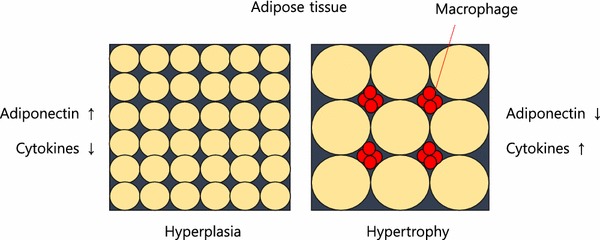
There are many causes of IR. Stress hormones (cortisol, adrenaline), inflammation, and high insulin levels themselves (response to dietary sugar and refined carbohydrates), each alone and in combination, cause immediate (within minutes to hours) insulin resistance. When these conditions persist over time insulin resistance becomes a chronic state. As fat cells grow in size, they reach a point where there is inadequate blood flow to the cells themselves and macrophages (immune cells that reside between the fat cells, most prominently in visceral fat) produce inflammatory chemicals called cytokines. Cytokines flow through the blood stream and effect every organ and every cell in the body creating a state of chronic inflammation which further worsens IR, creating a vicious cycle. As IR continues the pancreas produces increasingly higher amounts of insulin to maintain normal blood sugar levels but eventually IR becomes so great that blood sugar levels move into the “pre-diabetes” and eventually the diabetes range. IR builds for years to decades before blood sugar regulation fails. By the time blood sugar levels are “abnormal” insulin resistance has done great damage throughout the body.

Most doctors tragically do not order fasting insulin levels as routine blood tests. Fasting insulin levels rise long before fasting blood sugars and hemoglobin A1c start to rise. Meanwhile the damage progresses under the radar of routine testing.


1. Metabolic Engines: Muscle and Liver

Muscle

  • Normal Action: Insulin acts as a key that opens “doors” (GLUT4 receptors) to let glucose in for fuel. it also stimulates protein synthesis. Protein synthesis is essential to maintaining and increasing muscle mass and strength.
  • Insulin Resistance Effect: The “doors” stay locked. Glucose stays in the blood, and the muscle becomes “starched,” leading to sarcopenia (muscle wasting) and fatigue. The muscle can no longer utilize dietary protein to maintain or increase muscle mass.

Liver

  • Normal Action: Tells the liver to stop producing glucose and start storing it as glycogen or converting excess into fat.
  • Insulin Resistance Effect: The liver ignores the “stop” signal and keeps pumping out glucose while simultaneously ramping up fat production. This results in Non-Alcoholic Fatty Liver Disease (NAFLD).

2. Fat Cells (Adipose Tissue)

Visceral (Deep Fat) vs. Subcutaneous (Under Skin)

  • Normal Action: Insulin promotes fat storage and inhibits the breakdown of stored fat (lipolysis).
  • Insulin Resistance Effect: Fat cells—especially visceral ones—become “leaky.” They spill free fatty acids into the bloodstream and release inflammatory cytokines. This causes weight gain that is biologically difficult to lose because high insulin levels keep the “fat-burning” switch permanently off.

3. The Vital Organs: Heart, Kidneys, and Arteries

Heart and Arteries

  • Normal Action: Insulin stimulates the release of nitric oxide, which helps arteries relax and dilate.
  • Insulin Resistance Effect: Nitric oxide production drops, causing arteries to stiffen (hypertension). High insulin also damages the endothelial lining, leading to atherosclerosis (plaque buildup). This is the primary driver of heart failure, heart attacks and strokes.

Kidneys

  • Normal Action: Helps regulate sodium reabsorption.
  • Insulin Resistance Effect: The kidneys hold onto too much salt, increasing blood pressure. Over time, high blood sugar and inflammation damage the filtering units, leading to chronic kidney disease (CKD).

4. The Brain, Memory, and Sleep

Brain and Memory

  • Normal Action: Insulin crosses the blood-brain barrier to regulate appetite and support synaptic plasticity (the basis of learning).
  • Insulin Resistance Effect: Often called “Type 3 Diabetes,” brain IR starves neurons of energy and allows amyloid plaques and neurofibrillary tangles to build up. Worse, the brain is unable to utilize glucose to meet energy demands it starts to malfunction. This is a direct pathway to Alzheimer’s disease and dementia. As the small arteries in the brain become atherosclerotic and unable to deliver adequate oxygen and nourishment to brain cells small areas of the brain become permanently damaged eventually leading to vascular dementia.

Sleep

  • Insulin Resistance Effect: IR is heavily linked to Obstructive Sleep Apnea. (OSA) High insulin affects the central respiratory drive and increases fat deposits around the neck (a major contributor to obstructive sleep apnea), disrupting sleep cycles and creating periods of inadequate oxygen flow to the brain resulting in the acute stress response and awakening with each apneic event. Even without OSA, high insulin levels impair the production of melatonin which is essential to normal-restorative sleep. Throughout the day the brain accumulates metabolic toxins that must be cleared through the glymphatic system at night during sleep. As sleep is impaired this clearance system is disrupted, contributing to structural damage and functional loss. Sleep disruption and apneic episodes are stressful events, increasing stress hormones which then worsen IR, creating another vicious cycle. One night of sleep disruption causes acute IR. Chronic sleep disruption contributes to chronic IR.

5. Immunity and Structural Health

Immune System

  • Action: High insulin/glucose impairs white blood cell function.
  • Effect: Chronic inflammation (high CRP levels) and a weakened defense against infections. This is why diabetics often have poor wound healing. As normal immune regulation is impaired the immune system both over-reacts and under-reacts. Under-reaction increases risk of infection. Over-reaction produces cytokine storms seen with Covid-19 and other infections. Chronic inflammation worsens IR creating another vicious cycle. Chronic inflammation contributes to most chronic diseases.

Bone and Joints

  • Action: Insulin is bone-building.
  • Effect: IR leads to poor bone quality (despite high density) and osteoarthritis due to systemic inflammation and the “glycosylation” (sugar-coating) of joint cartilage, making it brittle.

6. The Pancreas: Beta and Alpha Cells

  • Normal Action: Beta cells produce insulin; Alpha cells produce glucagon (which raises sugar). They balance each other.
  • Insulin Resistance Effect:
    • Beta Cells: Work overtime to produce massive amounts of insulin to compensate, eventually “burning out” and dying. This can produce per4manent irreversible damage to the pancreas.
    • Alpha Cells: Become resistant to insulin’s “stop” signal and keep secreting glucagon, further raising blood sugar levels which in turn cause higher insulin secretion, both of which worsen IR, creating another vicious cycle.

7. Reproductive Effects: Infertility

  • In Women: High insulin stimulates the ovaries to produce excess testosterone, which is the primary driver of Polycystic Ovary Syndrome (PCOS) and infertility.
  • In Men: IR is a leading cause of low testosterone and erectile dysfunction (due to the arterial damage mentioned above).

Summary of Systemic Effects

ConditionPrimary Mechanism of Insulin Resistance
AtherosclerosisEndothelial dysfunction, high triglycerides, low HDL, increased TG/HDL ratio, increased small dense LDL and remnant particles, increased endothelial permeability.
DementiaNeuronal glucose starvation and plaque buildup, brain small vessel disease, disruption of blood brain barrier.
Chronic InflammationRelease of cytokines from visceral fat.
Heart FailureStiffening of the heart muscle and high blood pressure.
DiabetesPancreatic beta cell and alpha cell damage

Insulin’s Role vs. Insulin Resistance (IR)

Organ/SystemNormal Insulin ActionEffects of Insulin Resistance
LiverStops glucose production; stores glucose as glycogen.The liver ignores the “stop” signal, pumping out sugar even when you haven’t eaten (fatty liver).Fatty liver disease is the greatest cause of liver failure in the US.
MusclePrimary site for glucose uptake; promotes protein synthesis.Muscles can’t take in fuel efficiently, leading to fatigue and muscle wasting (sarcopenia). Muscle cells cannot use amino acids from dietary protein to maintain or build muscle. Elderly lose muscle and strength, resulting in falls, fractures and head trauma. Loss of muscle (the major sink for blood sugar after a meal) further increases duration and degree of blood sugar and insulin rise after a meal, which in turn increases IR. (vicious cycle)
Fat (Adipose)Stores fat; inhibits the breakdown of stored fat.Fat cells leak fatty acids into the blood, leading to high triglycerides and visceral fat gain. Macrophages (immune cells) produce inflammatory cytokines which circulate through the body contributing to chronic inflammation which worsens IR, another vicious cycle.
BrainRegulates appetite, memory, and cognitive function.Linked to “Type 3 Diabetes”; impaired memory and increased risk of neurodegeneration. Brain loses ability to meet energy demands and clear toxins. Insulin resistance in the brain explains memory loss, cognitive impairment, loss of neurons and synapses, loss of neuroplasticity. BDNF (brain derived neurotrophic factor) production is decreased by IR.
ArteriesStimulates nitric oxide for vasodilation (keeps vessels flexible).Reduced nitric oxide causes vessels to stiffen, raising blood pressure and plaque buildup. This is called endothelial dysfunction, the precursor to heart attack, stroke, peripheral vascular disease and a root cause for neuropathy and amputations.
HeartRegulates fuel use (switching between glucose and fats).The heart becomes “metabolically inflexible,” increasing the risk of heart failure.
KidneyManages sodium reabsorption and filtration.High insulin causes the kidneys to hold onto salt, driving up blood pressure and damaging filters. Oxidative stress leads to kidney failure.
Immune SystemModulates inflammation and helps T-cell function.Creates a state of “chronic low-grade inflammation” and weakens the response to infections.
BoneStimulates bone-forming cells (osteoblasts).Bone quality decreases; despite higher bone density in some cases, the bones are more brittle.
JointsMaintains cartilage and reduces systemic inflammation.High insulin promotes pro-inflammatory cytokines, accelerating osteoarthritis and gout.

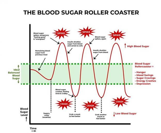
 A meal with sugar and refined carbohydrates causes excessive swings in blood sugar and insulin levels, creating insulin resistance and downstream damage. Alcohol consumption contributes to this process. Fat consumption does not cause a rise in blood sugar or insulin levels. Protein consumption produces a minimal rise in insulin levels in the absence of IR.

Fat storage can occur through hyperplasia (increase in number of fat cells) or hypertrophy (increase in size). Some ethnic groups are more prone to hypertrophy (south and east Asian). Hypertrophy in visceral fat (fat around the internal organs as opposed to fat under the skin) results in macrophage production of inflammatory cytokines. Eventually, the fat cells themselves can literally burst from too much volume.

 In my next post, I will discuss what we can do to prevent and reverse IR.

REFERENCES

Chadt A, Al-Hasani H. Glucose transporters in adipose tissue, liver, and skeletal muscle in metabolic health and disease. Pflugers Arch. 2020 Sep;472(9):1273-1298. doi: 10.1007/s00424-020-02417-x. Epub 2020 Jun 26. PMID: 32591906; PMCID: PMC7462924.

https://pmc.ncbi.nlm.nih.gov/articles/PMC7462924/

Fujita S, Rasmussen BB, Cadenas JG, Grady JJ, Volpi E. Effect of insulin on human skeletal muscle protein synthesis is modulated by insulin-induced changes in muscle blood flow and amino acid availability. Am J Physiol Endocrinol Metab. 2006 Oct;291(4):E745-54. doi: 10.1152/ajpendo.00271.2005. Epub 2006 May 16. PMID: 16705054; PMCID: PMC2804964.

https://pmc.ncbi.nlm.nih.gov/articles/PMC2804964

Vargas E, Joy NV, Carrillo Sepulveda MA. Biochemistry, Insulin Metabolic Effects. [Updated 2022 Sep 26]. In: StatPearls [Internet]. Treasure Island (FL): StatPearls Publishing; 2025 Jan-. Available from: https://www.ncbi.nlm.nih.gov/books/NBK525983/

https://www.ncbi.nlm.nih.gov/books/NBK525983/

Bugianesi E, Moscatiello S, Ciaravella MF, Marchesini G. Insulin resistance in nonalcoholic fatty liver disease. Curr Pharm Des. 2010 Jun;16(17):1941-51. doi: 10.2174/138161210791208875. PMID: 20370677.

https://pubmed.ncbi.nlm.nih.gov/20370677/

Cardillo C, Nambi SS, Kilcoyne CM, Choucair WK, Katz A, Quon MJ, Panza JA. Insulin stimulates both endothelin and nitric oxide activity in the human forearm. Circulation. 1999 Aug 24;100(8):820-5. doi: 10.1161/01.cir.100.8.820. PMID: 10458717.

https://pubmed.ncbi.nlm.nih.gov/10458717/

Ke JF, Wang JW, Zhang ZH, Chen MY, Lu JX, Li LX. Insulin Therapy Is Associated With an Increased Risk of Carotid Plaque in Type 2 Diabetes: A Real-World Study. Front Cardiovasc Med. 2021 Feb 1;8:599545. doi: 10.3389/fcvm.2021.599545. PMID: 33598483; PMCID: PMC7882504.

https://pubmed.ncbi.nlm.nih.gov/33598483/

Brosolo G, Da Porto A, Bulfone L, Vacca A, Bertin N, Scandolin L, Catena C, Sechi LA. Insulin Resistance and High Blood Pressure: Mechanistic Insight on the Role of the Kidney. Biomedicines. 2022 Sep 23;10(10):2374. doi: 10.3390/biomedicines10102374. PMID: 36289636; PMCID: PMC9598512.

https://pubmed.ncbi.nlm.nih.gov/36289636/

Kumar M, Dev S, Khalid MU, Siddenthi SM, Noman M, John C, Akubuiro C, Haider A, Rani R, Kashif M, Varrassi G, Khatri M, Kumar S, Mohamad T. The Bidirectional Link Between Diabetes and Kidney Disease: Mechanisms and Management. Cureus. 2023 Sep 20;15(9):e45615. doi: 10.7759/cureus.45615. PMID: 37868469; PMCID: PMC10588295.

https://pmc.ncbi.nlm.nih.gov/articles/PMC10588295/

Banks WA, Owen JB, Erickson MA. Insulin in the brain: there and back again. Pharmacol Ther. 2012 Oct;136(1):82-93. doi: 10.1016/j.pharmthera.2012.07.006. Epub 2012 Jul 17. PMID: 22820012; PMCID: PMC4134675.

https://pubmed.ncbi.nlm.nih.gov/22820012/

Rahman MS, Hossain KS, Das S, Kundu S, Adegoke EO, Rahman MA, Hannan MA, Uddin MJ, Pang MG. Role of Insulin in Health and Disease: An Update. Int J Mol Sci. 2021 Jun 15;22(12):6403. doi: 10.3390/ijms22126403. PMID: 34203830; PMCID: PMC8232639.

https://pmc.ncbi.nlm.nih.gov/articles/PMC8232639/

Scherrer U, Sartori C. Insulin as a vascular and sympathoexcitatory hormone: implications for blood pressure regulation, insulin sensitivity, and cardiovascular morbidity. Circulation. 1997 Dec 2;96(11):4104-13. doi: 10.1161/01.cir.96.11.4104. PMID: 9403636.

https://pubmed.ncbi.nlm.nih.gov/9403636/

Affuso F, Micillo F, Fazio S. Insulin Resistance, a Risk Factor for Alzheimer’s Disease: Pathological Mechanisms and a New Proposal for a Preventive Therapeutic Approach. Biomedicines. 2024 Aug 19;12(8):1888. doi: 10.3390/biomedicines12081888. PMID: 39200352; PMCID: PMC11351221.

https://pmc.ncbi.nlm.nih.gov/articles/PMC11351221/

Park MH, Kim DH, Lee EK, Kim ND, Im DS, Lee J, Yu BP, Chung HY. Age-related inflammation and insulin resistance: a review of their intricate interdependency. Arch Pharm Res. 2014 Dec;37(12):1507-14. doi: 10.1007/s12272-014-0474-6. Epub 2014 Sep 20. PMID: 25239110; PMCID: PMC4246128.

https://pubmed.ncbi.nlm.nih.gov/25239110/

Hardy OT, Czech MP, Corvera S. What causes the insulin resistance underlying obesity? Curr Opin Endocrinol Diabetes Obes. 2012 Apr;19(2):81-7. doi: 10.1097/MED.0b013e3283514e13. PMID: 22327367; PMCID: PMC4038351.

https://pmc.ncbi.nlm.nih.gov/articles/PMC4038351/

THIS WEBSITE PROVIDES INFORMATION FOR EDUCATIONAL PURPOSES ONLY. CONSULT YOUR HEALTH CARE PROVIDER FOR MEDICAL ADVICE.

Eat clean, drink filtered water, love, laugh, exercise outdoors in a greenspace, get some morning sunlight, block the blue light before bed, engage in meaningful work, find a sense of purpose, spend time with those you love, AND sleep well tonight.

Doctor Bob
The new dietary guidelines, a major improvement

The new Dietary Guidelines for Americans (DGA) represent a major improvement over previous guidelines. The storm of criticism registered by some “experts” and quoted by the media is NOT based on valid evidence. There has been excessive and irrational criticism over inverting the food pyramid, with animal sources of protein on the top. Until now recommendations for daily protein intake have been based upon estimates to avoid protein deficiency rather than optimization. The recommendations for 1.2-1.6 grams per kilogram bodyweight per day (up from 0.8 g/kg/day) is supported by data on muscle building, muscle maintenance and bone health (when combined with resistance exercise), especially for elderly individuals.

 Unfortunately, the 10% restriction on saturated fat remains, but the guidelines appropriately state that there is a lack of evidence to keep that restriction at 10% of caloric intake. The controversy over saturated fat remains despite meta-analyses of randomized controlled clinical trials that conclude that restriction of saturated fat has not been found to improve cardiovascular, metabolic, or cancer outcomes.

I doubt that those critical of the new DGA have read the 416-page appendices that document scientific references and explanations for each of the dietary recommendations. That’s right, 416 pages of narrative and scientific references are in the appendices of the DGA.

If you go here, you can download 4 documents including the Scientific Foundation Appendices (416 pages), the Scientific Foundation for DGA (90 pages), The Daily Servings Guide (3 pages) and the DGA (10 pages)

Here is an excerpt that is very useful.

How can you identify highly processed foods? Highly processed foods tend to have: 1. Refined grains and/or added sugars 2. Refined fats and oils 3. Long, complicated ingredient lists including chemical additives (e.g., artificial sweeteners, flavor enhancers, artificial colors, and emulsifiers). Examples are provided in Figures 4.3 and 5.8.

And a good explanation for why refined grains (any food made with flour) causes rapid and high blood sugar responses.

Refined Grains and Starches are Sugar • Refined grains are highly purified sources of starch. • Starches are long chains of glucose—a form of sugar. • During chewing and digestion, enzymes rapidly break down starch into glucose, raising blood sugar much like table sugar does. • Refined grain foods—white bread, crackers, breakfast cereals, chips, pastries, and pasta—can therefore act metabolically like sugar, delivering fast-absorbing carbohydrates with few nutrients or fiber to slow absorption. Take-home message: Refined grains are sugar in disguise. Choose whole grains, beans, or vegetables instead.

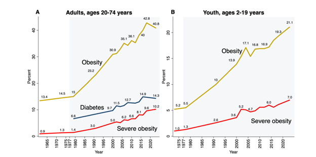
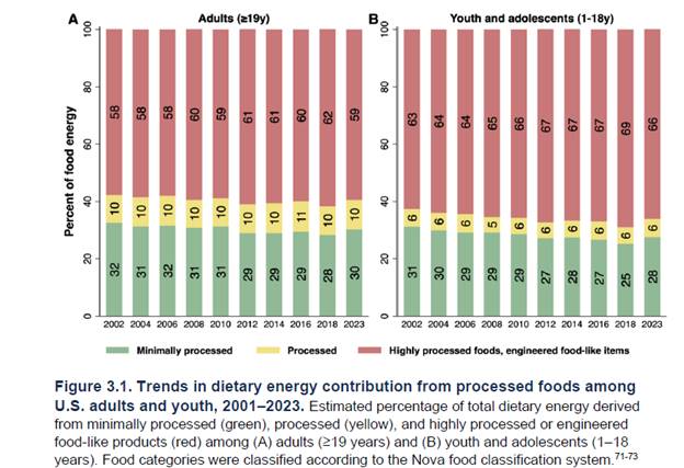
The following two graphics demonstrate the results of previous dietary guidelines that demonized healthy fats and animal sources of protein. The first shows the rise in obesity and diabetes but does not address the insulin resistance that evolves over decades before crossing the arbitrary threshold of diabetes, wreaking metabolic havoc long before diabetes occurs. The second graphic reveals how Americans consume 60% or more caloric intake in the form of refined carbohydrates (equivalent to sugar)

The following graphic displays the difference between minimally processed, moderately processed and highly processed foods according to the NOVA classification system (a system I will discuss and criticize in future posts). For now, suffice it to say that a simpler and more practical definition of “processed food” would include any food with one or more of the following: added sugar, artificial sweeteners, refined starch especially flour made from grains, refined “vegetable” oils, emulsifiers, artificial coloring, preservatives, and other additives found to disrupt the microbiome, intestinal barrier function or cause cancer. This graphic importantly covers the issue of food packaging which can contribute to the consumption of micro plastics, phthalates and PFAs

I have consistently advocated for a diet that consists of free-range meat, poultry, eggs, seafood (low mercury varieties), organic vegetables, fruits, nuts, seeds, an ancestral or paleo diet. The new DGA go a long way to produce evidence-based recommendations to help Americans eat healthy food. I continue to advocate for getting fiber from vegetables and fruits, eliminating grains, for reasons previously discussed but if you want to eat grains and are not gluten sensitive or suffer from celiac disease, consume the grains as a whole food, not in a food made from flour. “Whole grain bread” is not a whole grain food, nor is “whole grain” pasta. Once flour is made from grains the cellular components are destroyed producing a product with a glycemic index akin to sugar with the resultant metabolic disturbance which over the long run leads to insulin resistance, obesity and chronic disease.

If you want to consume dairy it makes much more sense to consume full fat fermented dairy foods instead of the low-fat dairy products advocated by prior DGA. The new DGA go into great detail to describe the nutrient deficiencies associated with vegan and vegetarian diets unless specific supplements are consumed. Specific recommendations for pregnant and breast-feeding mothers cover most important points as do age specific recommendations for infants and children. The importance of choline could have received a little more attention (best sources include eggs and liver). The importance of marine omega-3 fats (EPA, DHA, DPA) received adequate attention.

Overall, I consider the 2026 DGA a major improvement compared to previous iterations which ignored a large body of nutritional science.

The following references support my position on SFA and properly raised and prepared animal protein.

https://pubmed.ncbi.nlm.nih.gov/32562735/

Astrup A, Magkos F, Bier DM, Brenna JT, de Oliveira Otto MC, Hill JO, King JC, Mente A, Ordovas JM, Volek JS, Yusuf S, Krauss RM. Saturated Fats and Health: A Reassessment and Proposal for Food-Based Recommendations: JACC State-of-the-Art Review. J Am Coll Cardiol. 2020 Aug 18;76(7):844-857. doi: 10.1016/j.jacc.2020.05.077. Epub 2020 Jun 17. PMID: 32562735.

Reimara Valk, James Hammill, Jonas Grip, Saturated fat: villain and bogeyman in the development of cardiovascular disease?, European Journal of Preventive Cardiology, Volume 29, Issue 18, December 2022, Pages 2312–2321, https://doi.org/10.1093/eurjpc/zwac194

Associations of fats and carbohydrate intake with cardiovascular disease and mortality in 18 countries from five continents (PURE): a prospective cohort study Dehghan, MahshidDiaz, R et al. The Lancet, Volume 390, Issue 10107, 2050 – 2062

https://pubmed.ncbi.nlm.nih.gov/36216940/

Lescinsky H, Afshin A, Ashbaugh C, Bisignano C, Brauer M, Ferrara G, Hay SI, He J, Iannucci V, Marczak LB, McLaughlin SA, Mullany EC, Parent MC, Serfes AL, Sorensen RJD, Aravkin AY, Zheng P, Murray CJL. Health effects associated with consumption of unprocessed red meat: a Burden of Proof study. Nat Med. 2022 Oct;28(10):2075-2082. doi: 10.1038/s41591-022-01968-z. Epub 2022 Oct 10. PMID: 36216940; PMCID: PMC9556326.

Red and processed meat consumption and risk of incident coronary heart disease, stroke, and diabetes mellitus: a systematic review and meta-analysis Renata Micha1Sarah K WallaceDariush Mozaffarian, Circulation CIRCULATIONAHA.109.924977. Epub 2010 May 17. https://pubmed.ncbi.nlm.nih.gov/20479151/

Unprocessed Red Meat and Processed Meat Consumption: Dietary Guideline Recommendations from the Nutritional Recommendations (NutriRECS) Consortium Bradley C. Johnston, PhD, Dena Zeraatkar, Msc, et. al. Ann Intern Med 2019: 1:756-764 doi: 10.7326/M19-1621

Reduction of Red and Processed Meat Intake and Cancer Mortality and Incidence A Systematic Review and Meta-analysis of Cohort Studies Mi Ah Han, MD, PhD; Dena Zeraatkar, MSc; et. al., Ann Intern Med. 2019;171:711-720. doi:10.7326/M19-0699

Patterns of Red and Processed Meat Consumption and Risk for Cardiometabolic and Cancer Outcomes A Systematic Review and Meta-analysis of Cohort Studies Robin W.M. Vernooij, PhD*; Dena Zeraatkar, MSc Ann Intern Med.2019;171:732-741. doi:10.7326/M19-1583

Red and Processed Meat Consumption and Risk for All-Cause Mortality and Cardiometabolic OutcomesA Systematic Review and Meta-analysis of Cohort Studies Dena Zeraatkar, MSc, Mi Ah Han MD, PhD, et. al, Annals of Internal Medicine 1 October 2019: 171-710 doi: 10.7326/M19-0655

Effect of Lower Versus Higher Red Meat Intake on Cardiometabolic and Cancer Outcomes

A Systematic Review of Randomized Trials Dena Zeraatkar, MSc, Bradley C Johnston, PhD, et. al. Ann Intern Med. 2019;171:721-731. doi:10.7326/M19-https://doi.org/10.7326/M19-0622

E, Lavie CJ, Hill JO. The Failure to Measure Dietary Intake Engendered a Fictional Discourse on Diet-Disease Relations. Front Nutr. 2018 Nov 13;5:105. doi: 10.3389/fnut.2018.00105. PMID: 30483510 Archer; PMCID: PMC6243202.

Archer E, Hand GA, Blair SN (2013) Validity of U.S. Nutritional Surveillance: National Health and Nutrition Examination Survey Caloric Energy Intake Data, 1971–2010. PLoS ONE 8(10): e76632. https://doi.org/10.1371/journal.pone.0076632

O’Connor, Lauren E., et al. “Effects of total red meat intake on glycemic control and inflammatory biomarkers: a meta-analysis of randomized controlled trials.” Advances in Nutrition 12.1 (2021): 115-127.

Kiani AK, Dhuli K, Donato K, Aquilanti B, Velluti V, Matera G, Iaconelli A, Connelly ST, Bellinato F, Gisondi P, Bertelli M. Main nutritional deficiencies. J Prev Med Hyg 2022;63(suppl.3):E93-E101.https://doi.org/10.15167/2421-4248/jpmh2022.63.2S3.2752

Bailey RL, West KP Jr, Black RE. The epidemiology of global micronutrient deficiencies. Ann Nutr Metab. 2015;66 Suppl 2:22-33. doi: 10.1159/000371618. Epub 2015 Jun 2. PMID: 26045325.

Global, regional and national burdens of common micronutrient deficiencies from 1990 to 2019: A secondary trend analysis based on the Global Burden of Disease 2019 study. Zu Han et. al., eClinicalMedicine, February 11, 2022 https://doi.org/10.1016/j.eclinm.2022.101299

Sean R. Lynch, Why Nutritional Iron Deficiency Persists as a Worldwide Problem, The Journal of Nutrition, Volume 141, Issue 4, April 2011, Pages 763S–768S, https://doi.org/10.3945/jn.110.130609

Meat supplementation improves growth, cognitive, and behavioral outcomes in Kenyan children , J Nutr. 2007 Apr;137(4):1119-23.

Animal source foods have a positive impact on the primary school test scores of Kenyan schoolchildren in a cluster-randomised, controlled feeding intervention trial – PubMed (nih.gov), Hulett JL, Weiss RE, Bwibo NO, Galal OM, Drorbaugh N, Neumann CG.Br J Nutr. 2014 Mar 14;111(5):875-86. doi: 10.1017/S0007114513003310. Epub 2013 Oct 30.PMID: 24168874 Clinical Trial.

Meat supplementation increases arm muscle area in Kenyan schoolchildren – PubMed (nih.gov), Br J Nutr. 2013 Apr 14;109(7):1230-40. doi: 10.1017/S0007114512003121. Epub 2012 Aug 2.PMID: 22856533 Clinical Trial.

School snacks decrease morbidity in Kenyan schoolchildren: a cluster randomized, controlled feeding intervention trial.

Neumann CG, Bwibo NO, Jiang L, Weiss RE.Public Health Nutr. 2013 Sep;16(9):1593-604. doi: 10.1017/S1368980013000876. Epub 2013 Mar 28.PMID: 23537728

Effects of animal source foods, with emphasis on milk, in the diet of children in low-income countries.

Allen LH, Dror DK.Nestle Nutr Workshop Ser Pediatr Program. 2011;67:113-30. doi: 10.1159/000325579. Epub 2011 Feb 16.PMID: 21335994

https://www.bmj.com/content/351/bmj.h4962

The scientific report guiding the US dietary guidelines: is it scientific?

BMJ 2015; 351 doi: https://doi.org/10.1136/bmj.h4962 (Published 23 September 2015)

https://pmc.ncbi.nlm.nih.gov/articles/PMC9794145/

Teicholz N. A short history of saturated fat: the making and unmaking of a scientific consensus. Curr Opin Endocrinol Diabetes Obes. 2023 Feb 1;30(1):65-71. doi: 10.1097/MED.0000000000000791. Epub 2022 Dec 8. Erratum in: Curr Opin Endocrinol Diabetes Obes. 2025 Aug 1;32(4):166. doi: 10.1097/01.med.0001118356.22843.75. PMID: 36477384; PMCID: PMC9794145.

THIS WEBSITE PROVIDES INFORMATION FOR EDUCATIONAL PURPOSES ONLY. CONSULT YOUR HEALTH CARE PROVIDER FOR MEDICAL ADVICE.

Eat clean, drink filtered water, love, laugh, exercise outdoors in a greenspace, get some morning sunlight, block the blue light before bed, engage in meaningful work, find a sense of purpose, spend time with those you love, AND sleep well tonight.

Doctor Bob
Omega-3 in your diet and supplements

The benefit of omega-3 supplementation has been debated in the cardiology and nutritional literature for many years. Most studies of supplementation have failed to measure tissue levels achieved and often used very low doses. But when tissue levels were measured, either in the serum or red blood cell membrane, the studies consistently demonstrated significant reductions in all-cause mortality and cardiovascular mortality associated with high levels of omega-3 fatty acids.

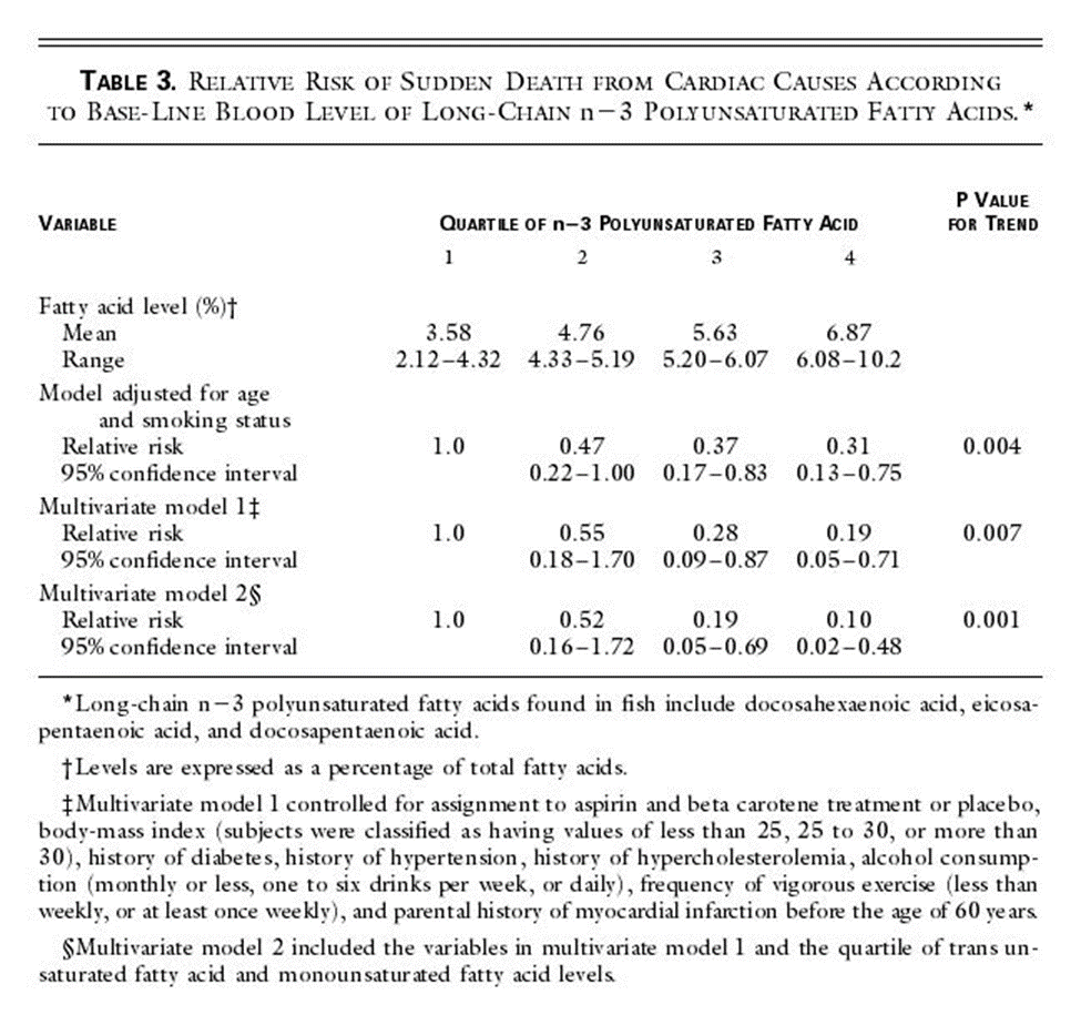
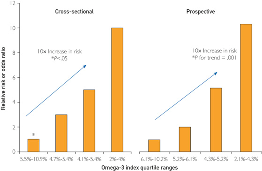
In addition, higher levels of omega 3 are associated with >=80% reduction in sudden death associated with acute myocardial infarction (acute MI) and > 80% reduction in sudden death in cohorts without known coronary artery disease followed long term.

Two Coronary CT Angiogram (CCTA) studies demonstrated that patients with stable coronary artery disease on statin therapy randomized to high dose EPA and DHA had “prevention of coronary plaque progression when an omega-3 fatty acid index >= 4% was achieved.”

 Another CCTA study demonstrated that patients receiving omega 3 supplementation had significantly less coronary atherosclerotic “high risk” lipid rich plaque prevalence (3.8% versus 32%) and lower total non-calcified plaque burden independent of cardiovascular risk factors compared to matched controls not receiving omega 3 supplements.

Omega 3 supplementation after an acute myocardial infarction has been found to reduce infarct size, reduce scaring (fibrosis), and enhance heart tissue healing. (Randomized controlled clinical trial) However a post MI study in 1027 elderly patients randomized to receive 1.8 grams per day of EPA+DHA versus a control group receiving corn oil showed no reduction in the primary composite cardiovascular endpoint between the two groups at 2 years but a higher incidence of AF in the omega 3 group that did not reach statistical significance.

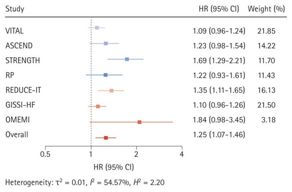
Recently a study, widely reported by the lay press, suggested that high dose omega-3 supplementation was associated with increased risk of atrial fibrillation (AF). These results conflicted with previous studies which demonstrated just the opposite, specifically prior studies demonstrated reduced risk of AF. The more recent study suffered a significant design flaw. The study in question failed to make statistical adjustment for the increased life span associated with higher levels of omega-3. Since age is a primary risk factor for AF, any intervention which increases life span would be expected to result in more AF over the lifetime of the patients as they aged (i.e., more elder years results in increased risk of AF). Therefore, statistical adjustment for that effect should be employed, but was not done in the study.

Unfortunately, science journalism has deteriorated to a state where the conclusions of study authors are most often quoted without interpretation or context, and without critical analysis or comparisons with previous studies that may have demonstrated opposing results.

In addition to large well-designed studies that have suggested a reduced risk of AF associated with omega-3 fatty acids, there have been natural experiments that provide reassuring information. The indigenous Inuit people of Greenland, for example, historically consumed large amounts of omega-3 in their diet with no evidence of increased risk of AF. In fact, before the introduction of western processed foods, estimates of AF among the Inuit were 0.6% (1963) compared to a “worldwide prevalence of AF in adults between 2 and 4%, between one and two percent in Canadian and the general US population and between 0.5% and 3% in most low- and middle-income countries.” A more recent study of Greenland yielded a prevalence of 1.4% likely reflecting a change in habits consisting of less exercise, more tobacco use and a shift to a more Western diet.

Still, multiple studies that used high dose omega 3 supplements in patients with known cardiac disease suggest an increased risk of AF. A good review of omega-3 fatty acids and atrial fibrillation was published in the Korean Journal of Internal Medicine, referenced below.

My interpretation of the complex data in this area is as follows.

At supplemental doses of EPA+DHA above 1.8 grams per day (and perhaps above 1 gram per day) in patients with known coronary artery disease (CAD), at high risk of CAD, or following a myocardial infarction, the risk of AF is increased by about 25% (relative risk). But the risk of lethal ventricular arrythmias (sudden death) associated with myocardial infarction (heart attack) is 80% lower in patients with a red blood cell omega 3 index of >=8. In people without known CAD, an omega-3 index >=8% is associated with an 80% reduction in sudden cardiac death. CCTA studies show significantly lower unstable “vulnerable” plaque in patients on omega-3 supplements. Similarly, omega 3 supplementation in patients on statins associates with halted plaque progression determined by serial CCTA in non-diabetics.

In addition, higher tissue levels of omega 3 are associated with significantly reduced all-cause, cardiovascular, and cancer mortality.

Omega-3 fatty acids are the chemical precursors of SPMs, specialized pro-resolving lipid mediators which help resolve inflammation. We know that cardiovascular events are driven by chronic inflammation in the walls of arteries, often mediated by insulin resistance. Chronic inflammation contributes to atherosclerosis (production of plaque in the artery wall) as well as cardiovascular events that result when unstable plaque ruptures.  Studies suggest that n-3 fatty acids may have antiarrhythmic properties with membrane-stabilizing effects in addition to antithrombotic and anti-inflammatory properties on the endothelial level. Basic science, observational studies and clinical trials have demonstrated that higher tissue levels of omega 3 fatty acids are associated with longer health span and lifespan. This understanding must be balanced with a probable increased risk of AF in certain clinical situations associated with high dose omega-3 supplements as described above (people with known CAD, high risk for CAD, or following and MI). Note that current AHA and ACC dietary guidelines include at least 2 servings of fatty fish per week, one serving provides approximately 1800 mg of omega-3.

Getting omega-3 fatty acids from cold water fatty fish would be ideal. Unfortunately, many individuals do not like salmon, sardines, mackerel or herring and simply will not consume enough of this fish to achieve protective tissue levels. Other species of fish and seafood provide much less amounts of omega 3. Another consideration is that individuals process omega 3 fats differently so different amounts of omega 3 will be necessary to reach the same protective levels in tissue. You can obtain a red blood cell omega-3 index using a home kit and a finger prick without a prescription (https://omegaquant.com/). The sample is mailed in to the lab and results reported directly to you. I have no financial relationship with these folks.

Bill Harris, PhD, is widely published in the area of omega-3 science. He developed the first clinically useful tissue assay which measures the % of omega 3 fat in red blood cell membranes, the “omega-3 index” which is the gold standard for omega 3 research and clinical testing. Although serum levels correlate with the red blood cell index, the later reveals dietary consequences of a 2-3 month period while serum levels reflect just a few days of most recent dietary habits. The red blood cell omega 3 index is analogous to the hemoglobin A1c which reveals average blood sugars over a 2–3-month period. Bill Harris suggests that 1800 mg per day of omega 3 fat consumption (food plus supplements) will achieve an index of >= 8% in most individuals.

Here are some references.

Harris WS, Tintle NL et.al., Fatty Acids and Outcomes Research Consortium (FORCE). Blood n-3 fatty acid levels and total and cause-specific mortality from 17 prospective studies. Nat Communications. 2021 Apr 22;12(1):2329. doi: 10.1038/s41467-021-22370-2. PMID: 33888689; PMCID: PMC8062567. https://pubmed.ncbi.nlm.nih.gov/33888689/

“Here we report the results of a de novo pooled analysis conducted with data from 17 prospective cohort studies examining the associations between blood omega-3 fatty acid levels and risk for all-cause mortality. Over a median of 16 years of follow-up, 15,720 deaths occurred among 42,466 individuals. We found that, after multivariable adjustment for relevant risk factors, risk for death from all causes was significantly lower (by 15-18%, at least p < 0.003) in the highest vs the lowest quintile for circulating long chain (20-22 carbon) omega-3 fatty acids (eicosapentaenoic, docosapentaenoic, and docosahexaenoic acids). Similar relationships were seen for death from cardiovascular disease, cancer and other causes”

Blood Levels of Long-Chain n–3 Fatty Acids and the Risk of Sudden Death Authors: Christine M. Albert, M.D., M.P.H., Hannia Campos, Ph.D., Meir J. Stampfer, M.D., Dr.P.H., Paul M. Ridker, M.D., M.P.H., JoAnn E. Manson, M.D., Dr.P.H., Walter C. Willett, M.D., Dr.P.H., and Jing Ma, M.D., Ph.D.

Published April 11, 2002 N Engl J Med 2002;346:1113-1118DOI:10.1056/NEJMoa012918 VOL. 346 NO. 15 https://www.nejm.org/doi/full/10.1056/NEJMoa012918

We conducted a prospective, nested case–control analysis among apparently healthy men who were followed for up to 17 years in the Physicians’ Health Study. The fatty-acid composition of previously collected blood was analyzed by gas–liquid chromatography for 94 men in whom sudden death occurred as the first manifestation of cardiovascular disease and for 184 controls matched with them for age and smoking status.

RESULTS

Base-line blood levels of long-chain n–3 fatty acids were inversely related to the risk of sudden death both before adjustment for potential confounders (P for trend = 0.004) and after such adjustment (P for trend = 0.007). As compared with men whose blood levels of long-chain n–3 fatty acids were in the lowest quartile, the relative risk of sudden death was significantly lower among men with levels in the third quartile (adjusted relative risk, 0.28; 95 percent confidence interval, 0.09 to 0.87) and the fourth quartile (adjusted relative risk, 0.19; 95 percent confidence interval, 0.05 to 0.71).

CONCLUSIONS

The n–3 fatty acids found in fish are strongly associated with a reduced risk of sudden death among men without evidence of prior cardiovascular disease.

Heydari B, Abdullah S, Pottala JV, Shah R, Abbasi S, Mandry D, Francis SA, Lumish H, Ghoshhajra BB, Hoffmann U, Appelbaum E, Feng JH, Blankstein R, Steigner M, McConnell JP, Harris W, Antman EM, Jerosch-Herold M, Kwong RY. Effect of Omega-3 Acid Ethyl Esters on Left Ventricular Remodeling After Acute Myocardial Infarction: The OMEGA-REMODEL Randomized Clinical Trial. Circulation. 2016 Aug 2;134(5):378-91. doi: 10.1161/CIRCULATIONAHA.115.019949. PMID: 27482002; PMCID: PMC4973577. https://www.ahajournals.org/doi/10.1161/CIRCULATIONAHA.115.019949

Conclusions: Treatment of patients with acute myocardial infarction with high-dose omega-3 fatty acids was associated with reduction of adverse left ventricular remodeling, noninfarct myocardial fibrosis, and serum biomarkers of systemic inflammation beyond current guideline-based standard of care.

Effect of Different Antilipidemic Agents and Diets on Mortality A Systematic Review

Studer M, Briel M, Leimenstoll B, Glass TR, Bucher HC. Effect of Different Antilipidemic Agents and Diets on Mortality: A Systematic Review. Arch Intern Med. 2005;165(7):725–730. doi:10.1001/archinte.165.7.725

Compared with control groups, risk ratios for cardiac mortality indicated benefit from statins (0.78; 95% CI, 0.72-0.84), resins (0.70; 95% CI, 0.50-0.99) and n-3 fatty acids (0.68; 95% CI, 0.52-0.90).

Feuchtner G, Langer C, Barbieri F, Beyer C, Dichtl W, Friedrich G, Schgoer W, Widmann G, Plank F. The effect of omega-3 fatty acids on coronary atherosclerosis quantified by coronary computed tomography angiography. Clin Nutr. 2021 Mar;40(3):1123-1129. doi: 10.1016/j.clnu.2020.07.016. Epub 2020 Jul 22. PMID: 32778459. https://pubmed.ncbi.nlm.nih.gov/32778459/

Conclusions: Omega-3-PUFA supplementation is associated with less coronary atherosclerotic “high-risk” plaque (lipid-rich) and lower total non-calcified plaque burden independent on cardiovascular risk factors. Our study supports direct anti-atherogenic effects of Omega-3-PUFA.

Alfaddagh A, Elajami TK, Saleh M, Mohebali D, Bistrian BR, Welty FK. An omega-3 fatty acid plasma index ≥4% prevents progression of coronary artery plaque in patients with coronary artery disease on statin treatment. Atherosclerosis. 2019 Jun;285:153-162. doi: 10.1016/j.atherosclerosis.2019.04.213. Epub 2019 Apr 13. PMID: 31055222; PMCID: PMC7963401.An omega-3 fatty acid plasma index ≥4% prevents progression of coronary artery plaque in patients with coronary artery disease on statin treatment – PMC (nih.gov)

Conclusions: EPA and DHA added to statins prevented coronary plaque progression in nondiabetic subjects with mean LDL-C <80 mg/dL, when an omega-3 index ≥4% was achieved. Low omega-3 index <3.43% identified nondiabetic subjects at risk of coronary plaque progression despite statin therapy

Association of Plasma Phospholipid Long-Chain Omega-3 FattyAcids with Incident Atrial Fibrillation in Older Adults: The Cardiovascular Health Study, Circulation Volume 125, Number 9 https://doi.org/10.1161/CIRCULATIONAHA.111.062653

Among 3326 US men and women ≥65 years of age and free of AF or heart failure at baseline, plasma phospholipid levels of eicosapentaenoic acid, docosapentaenoic acid, and docosahexaenoic acid were measured at baseline by use of standardized methods. Incident AF (789 cases) was identified prospectively from hospital discharge records and study visit ECGs during 31 169 person-years of follow-up (1992-2006).

Conclusions: In older adults, higher circulating total long-chain n-3 PUFA and docosahexaenoic acid levels were associated with lower risk of incident AF (atrial fibrillation). These results highlight the need to evaluate whether increased dietary intake of these fatty acids could be effective for the primary prevention of AF.

Omega-3 Fatty Acid Therapy: The Tide Turns for a Fish Story https://www.mayoclinicproceedings.org/article/S0025-6196(16)30764-9/fulltext

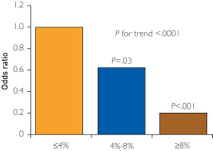
An omega-3 index of less than 4% is associated with increased CHD risk, particularly for sudden cardiac death. In contrast, an omega-3 index of more than 8% is associated with low CHD risk, whereas the range between 4% and 8% is considered intermediate risk

Risk of sudden death

Alfaddagh A, Elajami TK, Ashfaque H, Saleh M, Bistrian BR, Welty FK. Effect of Eicosapentaenoic and Docosahexaenoic Acids Added to Statin Therapy on Coronary Artery Plaque in Patients with Coronary Artery Disease: A Randomized Clinical Trial. J Am Heart Assoc. 2017; 6: e006981. 10.1161/JAHA.117.006981. https://pubmed.ncbi.nlm.nih.gov/29246960/

“High-dose eicosapentaenoic acid and docosahexaenoic acid provided additional benefit to statins in preventing progression of fibrous coronary plaque in subjects adherent to therapy with well-controlled low-density lipoprotein cholesterol levels.”

Huh JH, Jo SH. Omega-3 fatty acids and atrial fibrillation. Korean J Intern Med. 2023 May;38(3):282-289. doi: 10.3904/kjim.2022.266. Epub 2022 Dec 14. PMID: 36514212; PMCID: PMC10175873 https://pubmed.ncbi.nlm.nih.gov/36514212/

.

Effects of omega-3 fatty acid supplementation on the risk of atrial fibrillation. HR, hazard ratio; CI, confidence interval; VITAL, Vitamin D and Omega-3 Trial; ASCEND, A Study of Cardiovascular Events in Diabetes; STRENGTH, Statin Residual Risk with Epanova in High Cardiovascular Risk Patients with Hypertriglyceridemia; RP, Risk and Prevention Study; REDUCE-IT, Reduction of Cardiovascular Events With Icosapent Ethyl-Intervention Trial; GISSI-HF, Gruppo Italiano per lo Studio della Sopravvivenza nell’Insufficienza Cardiaca-Heart Failure; OMEMI, Omega-3 Fatty Acids in Elderly With Myocardial Infarction. Effects of omega-3 fatty acid supplementation on the risk of atrial fibrillation. HR, hazard ratio; CI, confidence interval; VITAL, Vitamin D and Omega-3 Trial; ASCEND, A Study of Cardiovascular Events in Diabetes; STRENGTH, Statin Residual Risk with Epanova in High Cardiovascular Risk Patients with Hypertriglyceridemia; RP, Risk and Prevention Study; REDUCE-IT, Reduction of Cardiovascular Events With Icosapent Ethyl-Intervention Trial; GISSI-HF, Gruppo Italiano per lo Studio della Sopravvivenza nell’Insufficienza Cardiaca-Heart Failure; OMEMI, Omega-3 Fatty Acids in Elderly With Myocardial Infarction. Effects of omega-3 fatty acid supplementation on the risk of atrial fibrillation. HR, hazard ratio; CI, confidence interval; VITAL, Vitamin D and Omega-3 Trial; ASCEND, A Study of Cardiovascular Events in Diabetes; STRENGTH, Statin Residual Risk with Epanova in High Cardiovascular Risk Patients with Hypertriglyceridemia; RP, Risk and Prevention Study; REDUCE-IT, Reduction of Cardiovascular Events With Icosapent Ethyl-Intervention Trial; GISSI-HF, Gruppo Italiano per lo Studio della Sopravvivenza nell’Insufficienza Cardiaca-Heart Failure; OMEMI, Omega-3 Fatty Acids in Elderly With Myocardial Infarction

THIS WEBSITE PROVIDES INFORMATION FOR EDUCATIONAL PURPOSES ONLY. CONSULT YOUR HEALTH CARE PROVIDER FOR MEDICAL ADVICE.

Eat clean, drink filtered water, love, laugh, exercise outdoors in a greenspace, get some morning sunlight, block the blue light before bed, engage in meaningful work, find a sense of purpose, spend time with those you love, AND sleep well tonight.

Doctor Bob
Paxlovid is a winner, but lifestyle changes are essential to mitigate Covid risk.

This discussion was originally posted before PAXLOVID WAS AVAILABLE. The title was originally Lifestyle More Effective than Drugs.

But now we have Paxlovid. Paxlovid is very effective in reducing morbidity and mortality associated with Covid-19 infection but also appears to reduce risk of Long Covid. Standard dosing: PAXLOVID two 150-mg tablets of nirmatrelvir, one 100-mg tablet of ritonavir twice daily for 5 days.

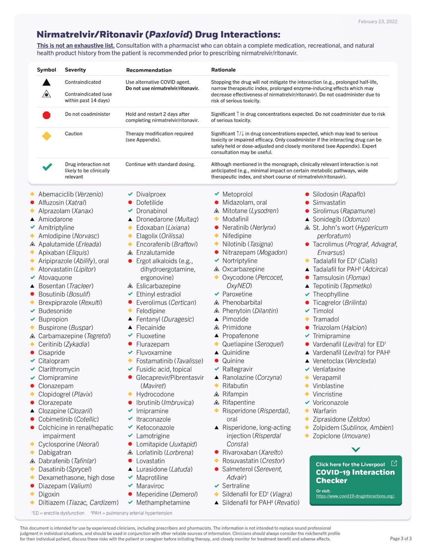
Dose adjustments are necessary for certain medical conditions and there are many drug interactions that should be considered. But do not let that discourage you from asking your provider to prescribe this drug for an acute Covid infection. This is a truly effective drug. It decreases morbidity and mortality as well as risk for Long Covid.

The results of a randomized placebo controlled clinical trial in high risk individuals has been published in the NEJM. The study was done just when Omicron hit. The study demonstrated an 89% reduction of hospitalizations and deaths by day 28 (absolute reduction of 6.2/100) with ZERO deaths in the Paxlovid group (7 in the placebo group). Paxlovid also had LESS side effects than placebo.

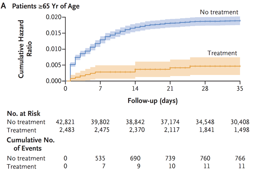
Another study from Israel demonstrated equally impressive results as shown here.

In addition, a study from the VA has looked at longer term effects (pre-print publication, still waiting for peer review.)

The study included 9000 Paxlovid patients treated within 5 days of symptom onset during the Omicron and subvariant waves and compared the treated patients with approximately 47,000 matched controls.

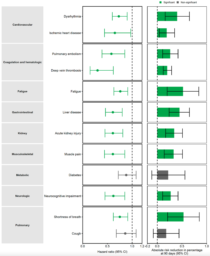
There was a 26% reduction in Long Covid.

Here is a breakdown of the Long Covid Symptoms

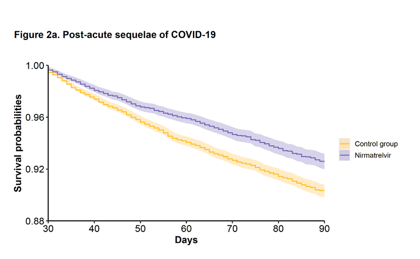
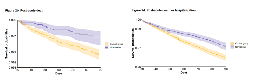
The VA study also showed a 48% reduction of death and 30% reduction in hospitalization after the acute phase (acute phase = first 30 days) as demonstrated here.

Many drug intervention trials for treating COVID-19 early in the pandemic have been disappointing. No studies have shown benefit for hydroxychloroquine, with or without azithromycin. This topic has been covered in previous posts. Remdesivir was FDA approved based upon one study that showed reduction in duration of symptoms. The mortality rate with Remdesivir, however, did not demonstrate a statistically significant difference when compared to “usual care”. https://www.niaid.nih.gov/news-events/nih-clinical-trial-shows-remdesivir-accelerates-recovery-advanced-covid-19

Another study published in Lancet failed to show any clinical benefit from Remdesivir.

“No statistically significant benefits were observed for remdesivir treatment beyond those of standard of care treatment. Our trial did not attain the predetermined sample size because the outbreak of COVID-19 was brought under control in China. Future studies of remdesivir, including earlier treatment in patients with COVID-19 and higher-dose regimens or in combination with other antivirals or SARS-CoV-2 neutralising antibodies in those with severe COVID-19 are needed to better understand its potential effectiveness.”

https://www.thelancet.com/journals/lancet/article/PIIS0140-6736(20)31022-9/fulltext

Likewise well designed studies of Ivermectin have shown no clinical benefit.

Monoclonal antibodies effective against early variants are no longer effective against the newer variants. So in terms of drug therapies for acute Covid infections we have Paxlovid for out patient care and dexamethasone for critically ill patients.

But we do know that certain underlying conditions such as obesity, diabetes, pre-diabetes (insulin resistance) and hypertension significantly increase risk of DEATH AND COMPLICATIONS with COVID-19. Since there are lifestyle interventions that can quickly and effectively mitigate these problems (diet, exercise, sleep, stress reduction….) now would seem like a good time to take our epidemics of obesity and diabetes in hand with aggressive lifestyle interventions to decrease the mortality rate of COVID-19 infection.

Such measures do not require expensive drugs or expensive drug trials, they simply require knowledge, guidelines and the will to implement change in our daily habits. Yet there has been little discussion about this in the media or on the part of public health officials.

Lets look at obesity in the US.

From 1999–2000 through 2017–2018, the age-adjusted prevalence of obesity increased from

30.5% to 42.4%, and the prevalence of severe obesity increased from 4.7% to 9.2%.

The most effective tool for addressing obesity and diabetes is a very low carbohydrate diet.

Effects of the Low Carbohydrate, High Fat Diet on Glycemic Control and Body Weight in Patients With Type 2 Diabetes: Experience From a Community-Based Cohort

https://pubmed.ncbi.nlm.nih.gov/32193200/

This study was a done in a community-based program, not an academic practice setting. That is significant since it demonstrates feasibility outside of academic centers with standard community resources. The results of this study confirmed the results of many previous studies done in academic settings including better blood sugar control, reduction or elimination of diabetic medications, and significant weight loss.

All patients following the LCHF diet who initially took
insulin had either a reduction or discontinuation of
this therapy by their healthcare provider when clinically
indicated, compared with less than a quarter of
those receiving usual care.

In another study done in Italy, significant weight reduction (7 kg), waistline reduction (7 cm.), fat mass reduction (3.8%) and systolic blood pressure reduction (10.5 mmHg) were achieved in 3 months with a Very Low Carbohydrate diet.

Middle and Long-Term Impact of a Very Low-Carbohydrate Ketogenic Diet on Cardiometabolic Factors: A Multi-Center, Cross-Sectional, Clinical Study (https://pubmed.ncbi.nlm.nih.gov/25986079/

Nina Teicholz had an opinion piece published in the Wall Street Journal on May 30, discussing the USDA dietary guidelines that have largely ignored a massive body of evidence supporting a Very Low Carbohydrate Diet for obesity and diabetes. She cites many studies that have been ignored by the USDA dietary guidelines committee. Here is here opening statement.

“Americans with obesity, diabetes, heart disease and other diet-related diseases are about three times more likely to suffer worsened outcomes from Covid-19, including death. Had we flattened the still-rising curves of these conditions, it’s quite possible that our fight against the virus would today look very different.”

You can read the full article here:

https://www.wsj.com/articles/a-low-carb-strategy-for-fighting-the-pandemics-toll-11590811260

But think about that simple statement, THREE TIMES MORE LIKELY TO SUFFER WORSENED OUTCOMES FROM COVID-19. Yet these conditions are highly responsive to lifestyle interventions that not only mitigate obesity, insulin resistance and high blood pressure, but also enhance immune function.

More from Teicholz’s opinion piece:

Other studies have found that dietary changes can rapidly and substantially improve cardiovascular risk factors, including conditions like hypertension that are major risk factors for worsened Covid-19 outcomes. A 2011 study in the journal Obesity on 300 clinic patients eating a very low-carbohydrate diet saw blood pressure quickly drop and remain low for years. And a 2014 trial on 148 subjects, funded by the National Institutes of Health, found a low-carb diet to be “more effective for weight loss and cardiovascular risk factor reduction” than a low-fat control diet at the end of the 1-year experiment.

In a recent letter to the editor published in the journal METABOLISM, Dr. Casey Means points out:

A diagnosis of diabetes has been a key indicator of the severity of
COVID-19, and in this regard, the virus has relentlessly highlighted our
global Achilles heel of metabolic dysfunction, and points to a prime opportunity
to fight back.
That fight, however, is not going to be won with Clorox, Purell,
masks, or anti-IL-6 drugs. The fight will only be won through a serious
commitment to improving everyone’s foundational metabolic health,
starting with the lowest hanging evidence-based fruit: dietary and lifestyle
interventions.

Read the full letter here: https://www.metabolismjournal.com/article/S0026-0495(20)30118-9/pdf

In 2 pages the letter describes multiple benefits of better glucose control relative to COVID -19 infection and the immune system as well as reduction of factors that lead to cytokine storm (terminal event for many COVID-19 patients). The letter also discusses the benefit of reducing environmental toxins (discussed in previous posts about COVID-19 and other health problems) that would likely benefit COVID-19 patients.

Research published April 18th, 2020 found that patients exposed to
highest amount of environmental nitrogen dioxide (NO2) had increased
risk of death fromCOVID-19, and that long-term exposure to this pollutant
may be one of the most important contributors to fatality by
compounding lung inflammation [20].

Minimizing exposure to environmental
pollutants may serve a role in quelling the underlying pro-inflammatory
state that characterizes metabolic disease and COVID-19 associated
cytokine storms
.

Other environmental toxins, including persistent organic pollutants
(POPs) found in air, water, and food generated from pesticides
and industrial chemicals, are also strongly implicated in the pathogenesis
of metabolic syndrome; promoting “clean living,” toxin-avoidant
strategies for patients as simple as emphasizing organic foods, home
air purification, and non-toxic home supplies could be considered, although
the clinical utility of these measures in the acute setting is unknown
[21].

In discussing the white elephant in the room he states:

What is starkly missing is the clear, simple, and strong recommendation for no added
sugar or ultra-refined carbohydrates, both of which are known drivers
of postprandial hyperglycemia and inflammation. As a medical community, we must not miss the opportunity to serve patients with straightforward, evidence-based nutritional and lifestyle strategies to assist in glycemic control.

I would encourage you to follow the link and read the 2 pages supported by multiple peer-reviewed references.

An ancestral (paleo) diet is also very effective for addressing insulin resistance, diabetes type 2 and obesity. Multiple studies have demonstrated this. Although an ancestral approach is typically low carb it is not typically ketogenic, but a ketogenic ancestral diet (high in non starchy vegetables to support the gut microbiome) can be implemented by restricting fruits to one serving of berries per day and limiting starchy vegetables.

Even without severe carbohydrate restriction, an ancestral anti-inflammatory diet will quickly address insulin resistance, type 2 diabetes, and obesity. In this study, insulin resistance was reversed in 10 days.

And another study compared an Ancestral (Paleo) diet to the Mediterranean diet in patients with ischemic heart disease AND insulin resistance (glucose intolerance or type 2 diabetes). Ancestral diet was superior to the Mediterranean diet in improving insulin sensitivity, blood sugar control and greater fat loss. Fasting blood sugars normalized in all patients on the Ancestral diet who had previously had abnormal blood sugars.

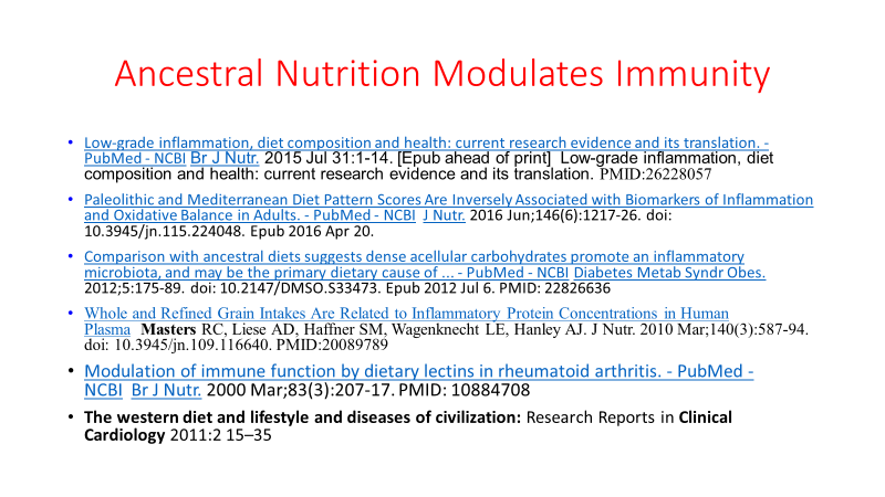
And here is a slide from one of my lectures with references on how an ancestral diet modulates immunity.

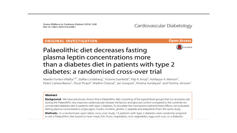
Leptin resistance, insulin resistance and obesity travel together. Here is yet another study demonstrating the effectiveness of an ancestral diet.

If you have obesity, diabetes or pre-diabetes the Very Low Carbohydrate version of the anti-inflammatory diet linked above would be the fastest and most effective intervention you can immediately employ to reduce your risk of succumbing to COVID-19. (Of course wear an N-95, follow good hygiene with hand-washing frequently, and use a HEPA filter or Corsi-Rosenthal box in your home, office, and enclosed work spaces)

In the context of the COVID 19 pandemic I will close with the usual summary.

  1. Avoid alcohol consumption (alcohol wreaks havoc with your immunity)
  2. Get plenty of sleep (without adequate sleep your immune system does not work well )
  3. Follow good sleep habits
  4. Exercise, especially out of doors in a green space, supports the immune system
  5. Get some sunshine and make sure you have adequate Vitamin D levels. Supplement with Vitamin D3 to get your levels above 30 ng/ml, >40ng/ml arguably better.
  6. Eat an anti-inflammatory diet rich in micronutrients.
  7. Practice stress reduction like meditation and yoga which improves the immune system
  8. Eliminate sugar-added foods and beverages from your diet. These increase inflammation, cause metabolic dysfunction, and suppress immunity.
  9. Eliminate refined-inflammatory “vegetable oils” from your diet, instead eat healthy fat.
  10. Clean up your home environment and minimize your family’s exposure to environmental toxins by following recommendations at EWG.org with regards to household products, personal care products, and organic foods. (https://www.ewg.org/)
  11. Drink water filtered through a high quality system that eliminates most environmental toxins.
  12. HEPA filters or the home-made version (Corsi-Rosenthal box) used in your home or workplace can reduce circulating viral load as discussed on this website.
  13. If you are eligible for vaccination, consider protecting yourself and your neighbor with a few jabs. Age > 50 and/or risk factors (Diabetes, pre-diabetes, insulin resistance, hypertension, obesity, heart disease, COPD, asthma, cancer treatment, immune suppression) suggests benefit from a booster. Risk for complications of boosters in adolescents, especially males, without risk factors, may equal benefit. Previous infection with Covid can be considered as protective as a booster. Discuss risk vs benefits with your doctor.

THIS WEBSITE PROVIDES INFORMATION FOR EDUCATIONAL PURPOSES ONLY. CONSULT YOUR HEALTH CARE PROVIDER FOR MEDICAL ADVICE.

Eat clean, drink filtered water, love, laugh, exercise outdoors in a greenspace, get some morning sunlight, block the blue light before bed, engage in meaningful work, find a sense of purpose, spend time with those you love, AND sleep well tonight.

Doctor Bob
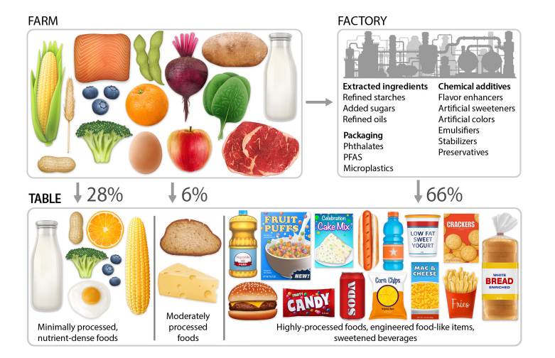
Obesity Epidemic Requires a Paradigm Shift

The obesity epidemic requires a paradigm shift. Several medical myths stand in the way of taking the most effective steps to safely help patients lose weight. The most important myth relates to saturated fat. Saturated fat consumption does not contribute to cardiovascular disease. This must be understood and accepted by the medical community so that sound advice can be given.

A meta-analysis of prospective epidemiologic studies showed that there is no significant evidence for concluding that dietary saturated fat is associated with an increased risk of CHD or CVD.( Am J Clin Nutr. 2010 Mar;91(3):497-9. )

In fact, as early as 2004, Mozaffarian et. al. investigated the influence of diet on atherosclerotic progression in postmenopausal women with quantitative angiography and found that:

In multivariate analyses, a higher saturated fat intake was associated with a smaller decline in mean minimal coronary diameter (P = 0.001) and less progression of coronary stenosis (P = 0.002) during follow-up. (Am J Clin Nutr. 2004 Nov;80(5):1175-84)

In addition, they further found that:

Carbohydrate intake was positively associated with atherosclerotic progression (P = 0.001), particularly when the glycemic index was high.

            Polyunsaturated fat intake was positively associated with progression when replacing other fats (P = 0.04)

These findings should come as no surprise given the basic science of atherosclerosis. Oxidized and glycated LDL stimulate macrophages to become foam cells initiating the creation of plaque. Cellular receptors that allow macrophages to ingest oxidized LDL are specific for oxidized LDL. These receptors do not recognize normal LDL to a significant degree.

Holovet et. al. studied the ability of oxidized LDL versus the Global Risk Factor Assessment Score (GRAS) to detect coronary artery disease. GRAS identified coronary artery disease 49% of the time, while oxidized LDL was correct 82% of the time.

In a large prospective study, Meisinger et al found that plasma oxidized LDL was the strongest predictor of CHD events when compared to conventional lipoprotein risk assessment and other risk factors for CHD.

Polyunsaturated fats are easily oxidized, saturated fats are not. It is the polyunsaturated fatty acids (PUFA) in the membrane of LDL particles that become oxidized and then initiate the cascade of inflammatory events leading to atherosclerosis. The major source of these PUFA in the American diet are “vegetable oils” (corn oil, soy oil etc.)  rich in the omega-6 PUFA, linoleic acid.

So why is this important to understand relative to the obesity epidemic? Because the most effective weight loss “diet” is arguably a low carbohydrate/high fat (LCHF) diet. This approach does not require calorie counting. This approach has been demonstrated to spontaneously reduce caloric intake whereas low fat diets require calorie counting and result in persistent hunger.

When compared to low fat calorie restricted diets  the LCHF approach has been equal or superior with respect to weight loss, insulin sensitivity, blood pressure reduction, and lipid profiles whenever these parameters have been measured.

But LCHF has not been embraced by the medical community due to the perceived dangers of saturated fat consumption and a low-fat ideology that lacks legitimate scientific evidence.

Once we dispel the mythology of saturated fat, the safety and efficacy of LCHF will be more readily accepted by physicians, the media and the lay public.

The nutritional villains in our society are highly refined and easily oxidized “vegetable oils” filled with pro-inflammatory omega-6 PUFA (linoleic acid), added sugar (especially HFCS) so prevalent in most processed foods and soft drinks, and the nutrient poor wasted calories of processed flour foods. These three culprits are responsible for our epidemics of obesity, insulin resistance and metabolic syndrome. These three conspire together to generate fatty liver disease, atherosclerotic plaque, and chronic inflammation.

When a LCHF approach is combined with  eating only fresh whole foods and avoiding added sugar, refined flour, and unhealthy  “vegetable oils”, we have the perfect recipe for our obesity epidemic.

The following references provide examples of studies that have demonstrated the efficacy, safety and  usual superiority of the LCHF  approach to weight loss.

Dig Dis Sci. 2007 Feb;52(2):589-93. Epub 2007 Jan 12. The effect of a low-carbohydrate, ketogenic diet on nonalcoholic fatty liver disease: a pilot study. Tendler D, Lin S, Yancy WS Jr, Mavropoulos J, Sylvestre P, Rockey DC Westman EC.

Fred Kummerow, PhD, fought the battle against Trans Fats for over 50 years.

Professor Fred Kummerow passed away on May 31 at his home in Urbana, Ill at age 102. He ate butter, red meat and eggs cooked in butter, along with plenty of fruits and vegetables. He avoided margarine, french fries and other fried foods, along with cookies, cake and crackers which contained artificial trans-fats. He conducted research in his nutrition science laboratory at the University of Illinois up until his death. he authored the book Cholesterol Won’t Kill You, But Trans Fat Could: Separating Scientific Fact from Nutritional Fiction in What You Eat

Fred warned the American public and scientists in the 1950s about the dangers of artificial man-made trans fats. His research was largely ignored and criticized by the food industry and by scientists who were funded by the food industry for decades. Despite mounting evidence in both animals and humans that artificial trans fats dramatically increased the risk of heart attacks, strokes, peripheral vascular disease, diabetes, obesity, and probably several forms of cancer, the FDA ignored his pleas to address the issue. In 2009 Professor Kummerow filed a petition with the FDA to ban the use of trans fats. Although federal law required that the FDA respond within 180 days to such a petition, the FDA remained silent. In 2013, approaching the age of 99, Professor Kummerow sued the FDA. Two years latter in 2015 the FDA declared that artificial trans-fats were unsafe and should be eliminated from the US food supply by 2018.

Through his lifelong work, Professor Kummerow has produced a policy change that will likely save hundreds of thousands of lives.

What are trans fats and why have they been in our food for 7 decades?

Although there are some forms of natural trans fats which are safe for consumption when consumed in whole foods, artificial trans-fats are produced by placing unsaturated fat (such as corn oil, soy oil) under high pressure and high temperature conditions and adding hydrogen in the presence of a metal catalyst. These fats were introduced to many American foods because they dramatically extend the shelf life of foods and give a pleasant mouth texture to a variety of processed foods. They remain in many foods still on the shelves today. You cannot rely on labels such as “NO TRANS FATS” OR “TRANS FAT FREE” because food companies are allowed to make this statement as long as the amount of trans fats does not exceed 0.5 grams per serving. No amount is safe!

The Institute of Medicine, on July 10, 2002 declared manufactured trans fatty acid (TFA) a serious danger to the health of our nation with a: “tolerable upper intake level of zero.”  This means there is no safe level of consumption. Despite that strong statement in 2002, it has taken the efforts of an elderly professor, including a lawsuit, to bring the FDA around to finally address the issue.

But it is not over yet, you can bet that the food industry will try to delay the implementation of the ban or possibly even argue against the overwhelming science that supports such a ban.

In the meantime read labels. If any food item contains “partially hydrogenated” oil of any kind or “hydrogenated oil” of any kind it contains trans fats. These foods are typically foods you should not be eating any way because they usually also contain added sugar, refined flour and/or refined easily oxidized inflammatory “vegetable” oils. They are not whole foods and therefore should not be consumed for many reasons. But if you want to eat cake, cookies, crackers, bread, or any other processed foods, beware and read the ingredients so as to at least avoid trans-fats.

You can read about Fred Kummerow, his life and research at these sites:

Fred A. Kummerow, scientist who raised early warnings about trans fats, dies at 102 – The Washington Post

Fred A. Kummerow, an Early Opponent of Trans Fats, Dies at 102 – The New York Times

Fred Kummerow, U. of I. professor who fought against trans fats, dies at 102 – Chicago Tribune

Fred also studied the effects of a oxysterols and oxidized low-density lipoprotein (OxLDL) both of which contribute to atherosclerosis.  In a  2013 publication Professor Kummerow stated

“levels of oxysterols and OxLDL increase primarily as a result of three diet or lifestyle factors: the consumption of oxysterols from commercially fried foods such as fried chicken, fish and french fries; oxidation of cholesterol in vivo driven by the consumption of excess polyunsaturated fatty acids from vegetable oils; and cigarette smoking.”

Yet the American Heart Association continues to recommend increased consumption of polyunsaturated fats from the likes of corn oil, soy oil, cottonseed and similar oils. I have discussed the problems with that advice here and here.

So the next time you avoid trans fats by reading food labels, think of Professor Kummerow who brought light to some very dark areas in the history of nutrition and food in the US.

Eat clean, live clean, and enjoy.

Dr. Bob

Sugar Industry paid Harvard researchers to trash fat and exonerate sugar!

By now most of you have already heard about the study published in JAMA that reveals an unsavory historical scenario wherein the sugar industry  funded an academic review paper that diverted the medical community’s attention from sugar as a vector for disease and erroneously placed it on saturated fat and cholesterol consumption. You can read about it by clicking on the following link.

How the Sugar Industry Shifted Blame to Fat – The New York Times

Here is a quote from the above cited article in the NY times:

The internal sugar industry documents, recently discovered by a researcher at the University of California, San Francisco, and published Monday in JAMA Internal Medicine, suggest that five decades of research into the role of nutrition and heart disease, including many of today’s dietary recommendations, may have been largely shaped by the sugar industry.

Here is the abstract of the article published in JAMA (Journal of the American Medical Association).

Sugar Industry and Coronary Heart Disease Research:  A Historical Analysis of Internal Industry Documents | JAMA Internal Medicine | JAMA Network

Early warning signals of the coronary heart disease (CHD) risk of sugar (sucrose) emerged in the 1950s. We examined Sugar Research Foundation (SRF) internal documents, historical reports, and statements relevant to early debates about the dietary causes of CHD and assembled findings chronologically into a narrative case study. The SRF sponsored its first CHD research project in 1965, a literature review published in the New England Journal of Medicine, which singled out fat and cholesterol as the dietary causes of CHD and downplayed evidence that sucrose consumption was also a risk factor. The SRF set the review’s objective, contributed articles for inclusion, and received drafts. The SRF’s funding and role was not disclosed. Together with other recent analyses of sugar industry documents, our findings suggest the industry sponsored a research program in the 1960s and 1970s that successfully cast doubt about the hazards of sucrose while promoting fat as the dietary culprit in CHD. Policymaking committees should consider giving less weight to food industry–funded studies and include mechanistic and animal studies as well as studies appraising the effect of added sugars on multiple CHD biomarkers and disease development.

This disturbing conspiracy reveals yet another industry sponsored distortion of science which had great impact on the health of our nation. The impact is accelerating today as the epidemics of obesity and diabetes rage out of control. But sugar consumption has not just been tied to obesity, diabetes, heart attacks and strokes. Sugar added foods and beverages have likely contributed to dementia,  many forms of cancer and other chronic debilitating diseases. Sugar and refined carbohydrates mediate these effects by increasing systemic inflammation and contributing to insulin resistance. Inflammation and insulin resistance are pathways to many disease processes. Metabolic syndrome (pre-diabetes) is the hallmark combination of multiple abnormalities with insulin resistance as the underlying root cause. Prolonged insulin resistance leads to type 2 diabetes and contributes to heart attacks, strokes,  cancer and dementia. In fact dementia is often referred to as type 3 diabetes, mediated in large part by insulin resistance in the brain.

Here are links to discussions and videos relevant to these topics.

Preventing Alzheimer’s Disease Is Easier Than You Think | Psychology Today

How to Diagnose, Prevent and Treat Insulin Resistance [Infographic] – Diagnosis:Diet

Reversing Type 2 diabetes starts with ignoring the guidelines | Sarah Hallberg | TEDxPurdueU – YouTube

I have previously provided links to the YouTube lectures given by the brilliant Dr. Jason Fung, These are worth mentioning again.

The Aetiology of Obesity Part 1 of 6: A New Hope

Insulin Toxicity and How to Cure Type 2 Diabetes

How to Reverse Type 2 Diabetes Naturally

Nina Teicholz is also worth a watch.

Nina Teicholz: The Big Fat Surprise – (08/07/2014)

And here is an important talk about sugar, refined carbohydrates and cancer.

Plenty to chew on.

We did not evolve to eat lots of sugar! It is dangerous stuff.

Bob Hansen MD


Berries, Dark Chocolate and Fruits-Vegetables improve blood vessels within 8 weeks

A recently published study reveals that within 8 weeks of consuming a combination of 70% dark chocolate (50 grams per day), one serving of berries, and four servings of polyphenol rich  fruits-vegetables per day, measurable improvements occur in the functioning of arteries in humans with high blood pressure. Just eight weeks and improvement was observed.

So what did the researchers do?

First, they took 102 adults with high blood pressure and instructed them to consume no chocolate, no berries and only 2 portions of fruits-vegetables per day for four weeks. After four weeks on a “low polyphenol diet” they measured forearm blood-flow response to two different vasodilators (a vasodilator increases blood flow by relaxing the muscle in the wall of arteries). Blood and urine samples were also taken.

After the four week period on a low polyphenol diet, 51 patients continued on the same diet (control group).  51 subjects had fruits and vegetables of their own choice (from a list rich in polyphenols), one serving of berries/day and 50 grams/day of 70% dark chocolate delivered free of charge to their homes weekly (intervention group).

All participants kept food diaries, answered food questionnaires, had regular consultations with nutritionists.

After 8 weeks, blood and urine testing demonstrated higher levels of polyphenols in the “high-polyphenol” group, as expected.

Most importantly the subjects consuming dark chocolate, berries and more vegetables-fruits on a daily basis demonstrated measurable and significant improvements in “endothelial function” compared to the other group. Endothelial function reveals the ability of arteries to respond to changes in demand for increased blood flow. “Endothelial” refers to the cells that line the walls of arteries, directly in contact with flowing blood.  These cells are endothelial cells.

The maximum forearm blood flow response to the infusion of acetyl-choline (a vasodilator) was the test used to measure endothelial function. The maximum forearm blood flow in the high polyphenol group was TWICE that of the low polyphenol group. This large difference occurred with just eight weeks of a simple dietary intervention.

Before the dietary intervention there was no measurable difference between the two groups of subjects. After the dietary intervention there was a very large and meaningful difference.

Endothelial dysfunction is a major predictor of cardiovascular risk (heart attack and stroke). Endothelial dysfunction is BAD.

You can read about polyphenols here .

Epidemiologic studies demonstrate the health benefits of diets rich in colorful vegetables and fruits. Among the fruits berries appear to have the greatest density of important micro-nutrients on a per calorie basis.

Dark chocolate has a high content of polyphenols and multiple studies suggest significant cardiovascular benefit with just an ounce or two per day. You must be careful though about your source since the dutch method of preparation depletes the polyphenols and some brands have high amounts of cadmium or lead. The richest source of dark chocolate identified by ConsumerLab with the lowest amounts of heavy metal contaminants is 100% (bitter) Bakers Dark Chocolate. Dip it in honey or have it with sweet berries to offset the bitter taste. Avoid in the evenings or late afternoon (the caffeine content can interfere with sleep)

So eat those colorful vegetables, berries and indulge in some dark chocolate.

Here is a suggested list of vegetables, Try to get 8-9 servings per day. I give this recommendation to my patients. I adopted this from the recommendations of Doctor Terry Wahls. (From a Wheelchair to Commuting on a Bicycle: How One Woman Naturally Reversed MS | Terry Wahls MD | Defeating Progressive Multiple Sclerosis without Drugs | MS Recovery | Food As Medicine)

9 SERVINGS  OF NON-STARCHY VEGETABLES PER DAY, 3 SERVINGS FROM EACH OF THREE CATEGORIES.


  1. DARK GREEN LEAFY VEGETABLES, 3 SERVINGS PER DAY EQUALS 3 CUPS MEASURED COOKED OR 6 CUPS MEASURED RAW
  • Arugula, Beet Greens, Bok Choy, Chard all colors, Chicory, Cilantro
  •  Dandelion Greens, Endive, Escarole, Kale-all kinds, Parsley, Radicchio
  • Radish leaves, Spinach, Turnip Greens, Watercress


  1. Colored vegetables, 3 cups daily:
  • GREEN: Artichoke, Asparagus, Avocado (FRUIT), Cabbage (red and green) Celery, Cucumber with skin, Okra, Olives, Peppers, Zucchini with skin
  • RED: Beets, red cabbage, red peppers, cooked tomatoes (fruit)
  • YELLOW: Carrots, Pumpkin, Squash-summer and winter, Sweet potato,


  1. SULFUR RICH VEGETABLES, 3 CUPS DAILY: Some leafy greens are also sulfur rich so there is overlap in these categories
  • Arugula, Asparagus, Bok Choy, Broccoli, Brussel sprouts, Cabbage, Cauliflower, Collard Greens, Garlic, Kale, Kholrabi, Leeks, Mushrooms, Onions red-yellow-white, Radishes, Scallions, Shallots, Turnip Greens, Watercress.

Eat the rainbow and enjoy good health.

You can read this study on Medscape but you must first establish a user name and password (free and open access)

Beneficial Effect of a Polyphenol-Rich Diet on CVD Risk

Bob Hansen MD.

Great lecture videos available on line

In January I attended the annual meeting of Physicians for Ancestral Health. There were great presentations on many topics related to lifestyle and health. Take a look at the website linked below to learn about many topics relating nutrition, exercise, and lifestyle to health.

Open Video Archives | Physicians for Ancestral Health

I presented a lecture titled “The Lipoprotein Retention Model, What’s Missing?” This discusses many factors (root causes) that contribute to the formation of plaque in arteries and what can be done to prevent this insidious process by adopting a “Paleo Lifestyle“.

Other videos include:

Paleopathology and the Origins of the Paleo Diet. Keynote speaker Michael Eades MD, author of several books and a well known website.

Medicine Without Evolution is like Engineering Without Physics– Randolph M Neese, MD Director of the Arizona State University Center for Evolution.

The Roles of Intermittent Fasting and Carbohydrates in Cancer Therapy– Dawn Lemanne, MD, MPH, integrative oncologist.

 23 and Me: Practical First Steps-Deborah Gordon MD, discusses a practical approach to utilizing information from this genetic test.

Histamine Intolerance-Why (food) Freshness Matters– Georgia Ede MD.


Mood and Memory: How Sugar Affects Brain Chemistry-Georgia Ede, MD.

Systems Analysis and Multiple Sclerosis– Tommy Wood MD, author, blogger and lecturer, frequently interviewed on topics related to exercise and nutrition.

Cholesterol OMG– Jeffry Gerber, MD “The Diet Doctor” in Denver Colorado

Bob Hansen MD