Category Archives: inflammation

Low Carb Beats Low Fat Again, Annals of Internal Medicine article

Once again, a randomized trial demonstrates that a carbohydrate restricted approach is superior to a low fat diet with regards to weight loss, inflammation, body composition and cardiovascular risk factors. This study was recently published in the Annals of Internal Medicine, the official journal for the American College of Physicians.

Men and women aged 22 to 75 years with a body mass index of 30 to 45 kg/m2 (obesity defined as BMI > 30, morbid obesity defined as BMI >35) were recruited from the general public by using mailing lists, fliers, work site and community screenings, and television advertisements.

Neither diet included a specific calorie or energy goal. Participants in each group were asked to refrain from changing their physical activity levels during the intervention

Here is the summary cut and pasted from the abstract.

Objective: To examine the effects of a low-carbohydrate diet compared with a low-fat diet on body weight and cardiovascular risk factors.

Design: A randomized, parallel-group trial. (ClinicalTrials.gov: NCT00609271)

Setting: A large academic medical center.

Participants: 148 men and women without clinical cardiovascular disease and diabetes.

Intervention: A low-carbohydrate (<40 g/d) or low-fat (<30% of daily energy intake from total fat [<7% saturated fat]) diet. Both groups received dietary counseling at regular intervals throughout the trial.

Measurements: Data on weight, cardiovascular risk factors, and dietary composition were collected at 0, 3, 6, and 12 months.

Results: Sixty participants (82%) in the low-fat group and 59 (79%) in the low-carbohydrate group completed the intervention. At 12 months, participants on the low-carbohydrate diet had greater decreases in weight (mean difference in change, −3.5 kg [95% CI, −5.6 to −1.4 kg]; P = 0.002), fat mass (mean difference in change, −1.5% [CI, −2.6% to −0.4%]; P = 0.011), ratio of total–high-density lipoprotein (HDL) cholesterol (mean difference in change, −0.44 [CI, −0.71 to −0.16]; P = 0.002), and triglyceride level (mean difference in change, −0.16 mmol/L [−14.1 mg/dL] [CI, −0.31 to −0.01 mmol/L {−27.4 to −0.8 mg/dL}]; P = 0.038) and greater increases in HDL cholesterol level (mean difference in change, 0.18 mmol/L [7.0 mg/dL] [CI, 0.08 to 0.28 mmol/L {3.0 to 11.0 mg/dL}]; P < 0.001) than those on the low-fat diet.

Limitation: Lack of clinical cardiovascular disease end points.

Conclusion: The low-carbohydrate diet was more effective for weight loss and cardiovascular risk factor reduction than the low-fat diet.

Primary Funding Source: National Institutes of Health.

Let’s go through those results again: At 12 months, participants on the low-carbohydrate diet had

  1.  greater decreases in weight. This has been demonstrated in multiple previously published studies.
  2.  greater decreases in  fat mass. This is an important distinction, the low carb group lost more fat, not muscle.
  3.  greater decreases in the ratio of total to high-density lipoprotein (HDL) cholesterol. This ratio is a measure of cardiovascular risk (risk for heart attack and stroke). It improved more on low carb than on low fat diets.
  4.  greater decreases in triglyceride level. Triglyceride level is also an important cardiovascular risk factor. It went down significantly more as compared to the low fat diet.
  5.  greater increases in HDL cholesterol level. This result is considered to be protective against heart attack and stroke.
  6. greater decreases in CRP level than those in the low-fat group. CRP (C-reactive protein) is a blood test for inflammation and is also a cardiovascular risk factor.
  7. significant decreases in estimated 10-year risk for coronary heart disease as measured by the Framingham risk analysis at 6 and 12 months, whereas those in the low-fat group did not. Say again, the low fat group did not decrease their Framingham risk analysis but the low carb group did.

All of these differences were “statistically significant”, meaning they were unlikely caused by accident.
And what about side-effects?

The number of participants who had symptoms, including constipation, fatigue, thirst, polyuria, diarrhea, heartburn, gas, nausea, vomiting, appetite changes, or headache, did not differ significantly between the low-carbohydrate and low-fat groups, except significantly more participants on the low-fat diet reported headaches at 3 months

The authors concluded:

Our study found that a low-carbohydrate diet induced greater weight loss and reductions in cardiovascular risk factors at 12 months than a low-fat diet among black and white obese adults who did not have diabetes, CVD, or kidney disease at baseline. Compared with a low-fat diet, a low-carbohydrate diet resulted in greater improvements in body composition, HDL cholesterol level, ratio of total–HDL cholesterol, triglyceride level, CRP level, and estimated 10-year CHD risk. Because CVD is the most common cause of death in the United States and obesity is a particularly prevalent risk factor, our study has important clinical and public health implications

Effects of Low-Carbohydrate and Low-Fat Diets: A Randomized Trial, A. Bazzano, MD, PhD, MPH et. al., Ann Intern Med. 2014;161(5):309-318. doi:10.7326/M14-0180

Get rid of the sugar-added foods, processed and refined flour foods and vegetable oils. Send a message to corporate America that crap-in-a bag and crap-in-a-box is no longer in demand. Eat only grass-fed meat, wild seafood, fresh vegetables, fresh fruit and tree nuts. Enjoy better health and better food.


Bob Hansen MD.

Chronic Pain Reduced by the Paleo Lifestyle

I spend 50% of my clinical time treating chronic pain patients. A paleolithic diet which consists of pastured grass-fed meat, free range poultry and eggs, fresh seafood, fresh vegetables, fruits and nuts decreases inflammation by eliminating major sources of dietary induced inflammation.

Yesterday I saw a patient one month after he started a paleolithic lifestyle (paleo diet, 8 hours of sleep per night- cycling with the sun, regular exercise including a prescribed spine rehab program).

Within 30 days his pain  has decreased by more than 50%, He feels  more energetic. He stated “I have started to dream again and get a full night’s sleep”. He has lost 12 pounds in one month and his blood pressure is down. He is ready to return to work after not working for eight months (with some activity restrictions). He is not taking any opiate pain medication.

His MRI scan and X-rays of the spine will not demonstrate any improvement. He still has degenerative disc disease, one or more tears in a disc annulus (outer wall of the disc) and arthritis in the facet joints of his neck (cervical spine) and lower back (lumbar spine). But the lifestyle elements that have contributed to his chronic inflammation have been significantly reduced in just 30 days and he has benefited “tremendously” in his own words.

There are many mechanisms involved with chronic inflammation. Most patients with chronic pain have an inflammatory component. Many patients with chronic pain are overweight or obese. Excess visceral adiposity (fat around the internal organs) creates a state of chronic inflammation by constantly producing inflammatory chemicals called chemokines and cytokines. These inflammatory mediators are produced by the fat cells and by the white blood cells (macrophages) that reside alongside the fat cells. They contribute to a process called central sensitization where the brain and spinal cord nerves that mediate pain  become sensitized and over-react to sensory input. Interleukin 6 is one of these mediators. Increased levels are associated with fatigue, depression and a state of hyperalgesia where painful stimuli are amplified. Tumor necrosis factor alpha is another important inflammatory mediator produced in excess when excess fat accumulates around the internal organs. Weight loss is essential to decease systemic inflammation, particularly in the setting of chronic pain when someone is overweight or obese.

Pro-inflammatory foods can also increase inflammation by altering intestinal flora and increasing intestinal permeability. These mechanisms have been discussed in previous posts and in the manifesto page of this website.

Few patients follow my dietary and lifestyle advice. Most seem to prefer taking pills, getting injections and other interventional pain procedures. In other words, they prefer to “be-fixed” rather than  take lifestyle initiatives that are likely to not only decrease their pain but also improve their general health. As an interventional pain practitioner I encourage patients to take full advantage of the pharmacology and interventional procedures that are likely to help. But without significant changes in bad dietary habits, poor sleep hygiene and without adopting a rehabilitation exercise program the pills and injections/procedures are much less effective and the prognosis is poor.

Stress reduction is also essential for health in general and for pain reduction in particular. Yet despite repeated recommendations to utilize an inexpensive stress reduction workbook, few patients ever bother to take this important step to reduce pain, anxiety and suffering.

Our culture is one in which patients expect to “be fixed” rather than to be led down a path which leads to healing and functional improvement by actively participating in their own rehabilitation and healing. Our culture is also one in which  major organizations provide bad dietary advice, particularly with respect to encouraging increased consumption of grains and legumes which have pro-inflammatory components and anti-nutrients. We evolved over a few million years without consuming grains, legumes, refined vegetable olis or dairy. Our evolutionary biology and physiology thrive when these foods, particularly processed foods are eliminated from the diet and we consume only those whole natural foods we have evolved to eat.

Modern medicine provides many remarkable drugs, surgeries and procedures that can be life saving and life altering. But application of this technology without addressing the fundamental determinants of health (proper nutrition, restorative sleep, judicious exercise, stress reduction, and restoration of circadian rhythm) yields much less benefit. Ultimately, unless we remove from our lives the destructive components of modern society and culture we cannot heal and instead continue to suffer from chronic degenerative diseases that cause pain, loss of intellect and loss of mobility.

No references tonight, just comments and reflection. References have been provided in previous posts.

Peace, health, and happiness.

Dr. Bob

The bacteria in your gut are essential to your health Part II, obesity, metabolic syndrome and dysbiosis

I have discussed the evidence linking the mix of bacteria in your gut (gut flora) to health and disease in Part I. The Bacteria in your Gut are essential to your health Part I | Practical Evolutionary Health

Today I will discuss the evidence related specifically to  obesity and metabolic syndrome (the constellation of obesity, insulin resistance, high blood pressure, and abnormal blood lipids). My discussion will follow closely the evidence and theory presented in research and review papers authored by Dr. Cani and colleagues. The first one is titled:

Gut microbiota controls adipose tissue expansion, gut barrier and glucose metabolism: novel insights into molecular targets and interventions using prebiotics.”

You can find the full text of this article here .

I have had the pleasure of corresponding with Dr. Cani by e-mail regarding her many publications investigating the relationship between gut flora, obesity, and metabolic syndrome.

“Recently, we and others have identified several mechanisms linking the gut microbiota with the development of obesity and associated disorders (e.g. insulin resistance, type 2 diabetes, hepatic steatosis).”

Explanation: The gut microbiota are the bacteria, viruses and other “bugs” that reside in our intestines. Insulin resistance can occur in various parts of the body, wherever insulin has an effect including fat cells, liver, muscle, brain. When higher amounts of insulin are required to achieve an effect this is called insulin resistance. In Type 2 diabetes, the pancreas is still able to make insulin but insulin is less effective in controlling blood sugar. In Type I diabetes the pancreas no longer produces insulin. Hepatic Steatosis means fatty liver disease. The liver accumulates fat and this can lead to cirrhosis, liver failure and death. Alcohol consumption can cause this but when alcohol is not involved this is called Non-Alcoholic-Fatty-Liver Disease (NAFLD). Our nation presently has an epidemic of not just obesity but also NAFLD. Evidence points to  excess carbohydrate consumption and excess consumption of vegetable oils (linoleic acid)  as contributing factors in NAFLD.  Carbohydrate restriction and consumption of saturated fat, particularly medium chain fats (as found in coconut) can protect against NAFLD. But the gut flora also play a role. The mechanisms involved are many.

“Among these, we described the concept of metabolic endotoxaemia (increase in plasma lipopolysaccharide levels) as one of the triggering factors leading to the development of metabolic inflammation and insulin resistance.”

Endotoxemia occurs when a toxin from certain kinds of bacteria circulates in the blood. This endotoxin enters our blood through our intestines under conditions in which the protective barrier of the intestines is compromised. The compromise of the intestinal barrier is variously referred to as ” leaky gut” or “increased intestinal permeability”. Wheat gluten-gliadin  causes increased intestinal permeability (especially in celiac disease) as can other plant lectins. In this discussion, the gut bacteria also contribute in the setting of “dysbiosis” (the beneficial effects of helpful bacteria are overwhelmed by the harm-causing bacteria when a healthy balance is not present)

Lipopolysaccharide (LPS) comes from the outer wall membrane of certain bacteria. Blood plasma is the liquid part of blood in which the blood cells circulate. So an “increase in plasma lipopolysaccharide” simply means that there is more LPS circulating in the blood. That is a bad thing. Depending on how much is circulating this alone can cause organ failure and death and is a major part of the physiologic changes involved in septic shock. But lower levels of LPS circulating in the blood can cause chronic low grade inflammation and insulin resistance. Obesity is associated with chronic inflammation and increased LPS circulating in the blood and being distributed to various organs where it wreaks havoc.

“Growing evidence suggests that gut microbes contribute to the onset of low-grade inflammation characterizing these metabolic disorders via mechanisms associated with gut barrier dysfunctions.”

“We have demonstrated that enteroendocrine cells (producing glucagon-like peptide-1, peptide YY and glucagon-like peptide-2) and the endocannabinoid system control gut permeability and metabolic endotoxaemia.”

That is a mouth-full. Over thirty different kinds of hormone producing cells have been found in the human intestine. These cells are called enteroendocrine cells. The hormones produced by these cells have many effects. You can find a great review of these cells and their effects here .

In Dr. Cani’s review article she describes how some of these hormones produced in the gut can increase intestinal permeability and allow more of the toxic, inflammation producing LPS to enter the bloodstream. But these hormonal effects are just part of the picture. Another part relates to endocannabinoids.

The  Endocannabinoid system in humans is complex and relates to hunger, satiety, energy metabolism, and yes gut permeability. Endocannabinoid refers to our internal (endo) production of cannabis like substances. Pot smoking people get the munchies because of the appetite stimulating effects of marijuana. But endocannabinoids have many other physiologic effects including the modulation of pain, mood, immune function and memory.

Dr. Cani describes in great detail the evidence supporting the roles that the gut flora play in influencing intestinal permeability mediated through the effects of various hormones and endocannabinoids. In animal and human studies changing the gut flora produces changes in these hormones and endocannabinoids which in turn can increase or decrease intestinal permeability and increase or decrease circulating LPS.

It turns out that specific  Prebiotics can produce growth of beneficial gut bacteria and through the series of steps outlined above, reduce inflammation in the body, improve blood sugar, improve insulin sensitivity, and decrease fat,

Oh, and similar to the endocannabinoid system, there is an “apelinergic system” in our bodies that also plays a role. If you want to read more about these systems you should read the original article and the other links below to related articles.

I have discussed in the past that fecal transplants have been used to treat the specific dysbiosis that occurs with C Difficile colitis. But fecal transplants have many potential beneficial uses.

The Fatlose 2 trial is presently studying the effects of fecal transplants on insulin resistance and related problems in human volunteers. I will let you know when the results are published, Studies conducted in rodents have demonstrated significant weight loss and improved insulin sensitivity when obese rodents receive fecal transplants from lean rodents.

In summary: dysbiosis represents an unhealthy mix of bacteria in the gut

  • dysbiosis causes increased intestinal permeability (leaky gut)
  • increased intestinal permeability leads to increased circulating LPS, which is bad
  • elevated levels of circulating LPS create a chronic state of inflammation which contributes to obesity and metabolic syndrome
  • the mechanisms that link dysbiosis to intestinal permeability include hormonal disruption (enteroendocrine cells) and the endocannabinoid system. Other mechanisms are also likely in play.
  • prebiotics and probiotics can mitigate dysbiosis, reduce intestinal permeability, reduce inflammation, and offer potential therapy for obesity and metabolic syndrome
  • fecal transplantation offers a potential for treatment for obesity and metabolic syndrome, research is underway

Our ancestors lived and evolved for a few million years prior to the relatively brief ten thousand years of agriculture and one hundred years of industrialization. The overuse of antibiotics in medicine and animal husbandry have contributed to dysbiosis. Other factors include stress, disruption of circadian rhythm, sleep deprivation. Cesarean delivery and avoidance of breast feeding conspire to dysbiosis. Processed foods feed unfriendly bacteria in our guts at the expense of beneficial bugs. Agricultural foods have introduced dietary lectins which also increase intestinal permeability and thereby contribute to chronic inflammation. The further we stray from our evolutionary niche, the more problems we experience.

This discussion just touches the surface of gut flora, dysbiosis, health and disease. We have yet to explore the gut-brain axis. Our gut and microflora communicate with and effect the function of our brain and other organs as well.

We will continue to explore health and disease from an evolutionary perspective.

Below are links to articles related to our discussion.

Peace, health and happiness.

BOB

Gut microbiota controls adipose tissue expans… [Benef Microbes. 2014] – PubMed – NCBI

Glucose metabolism: Focus on gut microbiota, … [Diabetes Metab. 2014] – PubMed – NCBI

Probiotics, prebiotics, and the host microb… [Ann N Y Acad Sci. 2013] – PubMed – NCBI

Crosstalk between the gut microbiota a… [Clin Microbiol Infect. 2012] – PubMed – NCBI

Gut microbiota and its possible relationship … [Mayo Clin Proc. 2008] – PubMed – NCBI

Enteroendocrine Cells: Neglected Players in Gastrointestinal Disorders?

The Bacteria in your Gut are essential to your health Part I

Our human body consists of about 100 trillion cells but we carry about 1000 trillion bacteria in our intestines, that represents 10 times the amount of our own cells. (1) These bacteria are variously called our micro-flora, microbiome, gut flora, etc, along with viruses and other organisms that co-exist and co-evolved with us. Advances in rapid gene identification have enabled an explosion of knowledge related to our micro-flora, health and disease. We each carry an estimated 500 to 1000 different species of bacteria in our intestines and the balance/mix of these bacterial species can have profoundly positive or negative affects on our health. Patterns of micro-flora have been identified for a variety of human disorders including obesity, diabetes type I, several kinds of cancer and  inflammatory bowel disease to name a few. The issue of association vs. causation remains to be resolved but the beneficial and therapeutic effects of pro-biotics and fecal transplant (in rodent and human studies) in a variety of situations along with the observed deleterious effects of interrupting our micro-flora speak in favor of a causative or contributory role. (2) (3)

Accumulating evidences indicate that some diseases are triggered by abnormalities of the gut microbiota. Among these, immune-related diseases can be the promising targets for probiotcs. Several studies have proved the efficacy of probiotics for preventing such diseases including cancers, infections, allergies, inflammatory bowel diseases and autoimmune diseases. Lactobacillus casei strain Shirota (LcS) is one of the most popular probiotics, benefits of which in health maintenance and disease control have been supported by several science-based evidences.(2)

Early microbial colonization of the gut reduces the incidence of infectious, inflammatory and autoimmune diseases. Recent population studies reveal that childhood hygiene is a significant risk factor for development of inflammatory bowel disease, thereby reinforcing the hygiene hypothesis and the potential importance of microbial colonization during early life. (3)

Early-life environment significantly affects both microbial composition of the adult gut and mucosal innate immune function. We observed that a microbiota dominated by lactobacilli may function to maintain mucosal immune homeostasis and limit pathogen colonization. (3)

The human GI tract starts with the mouth and ends with the rectum. In between lay the esophagus, stomach, and intestines which consist of the duodenum, jejunum, ileum, and colon.

The surface area of the intestines equals that of a tennis court providing a huge area for absorption, digestion and interaction between our immune system and the micro-flora. This large surface area is the result of the intestinal micro-villi which produce an undulating surface resembling a series of peaks and valleys. The constant interplay between our immune system (4) and our micro-flora from birth to death along with the signaling and communication that occurs between our micro-flora and our nervous system (5,6,7) present two physiologic mechanisms for potential symbiosis (mutually beneficial interaction) vs dysbiosis (disease causing relationship).

Before birth the mouth, skin and intestine of the fetus is sterile. The first major introduction of bacteria to the infant occurs with birth  when the infant swallows bacteria in the mother’s birth canal and the infant’s skin becomes colonized by the mother’s bacteria. Infants born by cesarean section lack this initial exposure and they suffer increased risk of allergic and auto-immune disease (8). The rate of cesarean section in the US is now about 30 % and along with that increase there has been an observed increase in allergy, auto-immune and other diseases.

The second major addition to human gut and skin flora occurs with breast feeding and again breast-fed infants show decreased rates of allergy and auto-immune disease as well as decreased infections compared to bottle fed infants.

The interaction between the micro-flora and the immune system presents many complex relationships and interactions. Immune tolerance allows the immune system to recognize “self” and “friendly bacteria”  limiting the development of auto-immune disease and enhancing anti-inflammatory processes. At the other extreme recognition of “non-self”  allows for the recognition and disposal of “foreign” invaders such as infections or mutated cancer cells.

“The Old Friends Hypothesis”
Common organisms interact with dendritic cells in the GI tract, leading to increased maturation of dendritic cells. When there is interaction with these organisms again, the dendritic cells increase Treg maturation; not Th1 or Th2. This increases the baseline amount of anti-inflammatory cytokines, producing a Bystander Suppression. Another consequence of the increased number of mature dendritic cells is as they interact with self antigens, they increase the number Treg specific to these antigens. This is referred to as Specific Suppression. Together these two arms lead to tolerance of both self antigens as well as those of helpful gut organisms. (8)

Translation:  Treg or Regulatory T cells regulate the immune system and help prevent auto-immune disease and allergic reactions. Th1 and Th2,  T helper cells , on the other hand, increase inflammation and help our bodies defend against infection. The balance between Tregs and Th1, Th2 cells governs inflammatory responses.

Premature infants have an increased risk of a developing a very severe illness called necrotizing enterocolitis. Human studies have demonstrated significant risk reduction for this problem with the administration of pro-biotics to infants in neonatal intensive care units. (9)

Similarly, administration of pro-biotics during the first few years of life (to mother and child)  have been associated with decreased risk of eczema in children. While some studies suggest reduction of allergies and asthma in children, the regular use of probiotics remains undecided relative to preventing food allergies or asthma (10, 11).

Due to the recent exponential increase in food allergies and atopic disorders, effective allergy prevention has become a public health priority in many developed regions. Important preventive strategies include the promotion of breastfeeding and vaginal deliveries, judicious use of perinatal antibiotics, as well as the avoidance of maternal tobacco smoking. Breastfeeding for at least 6 months and introduction of complementary solids from 4-6 months are generally recommended. Complex oligosaccharides in breast milk support the establishment of bifidobacteria in the neonatal gut which stimulate regulatory T lymphocyte responses and enhance tolerance development…Perinatal supplementation with probiotics and/or prebiotics may reduce the risk of atopic dermatitis, but no reliable effect on the prevention of food allergy or respiratory allergies has so far been found. A randomized trial on maternal fish oil supplementation during pregnancy found that atopic dermatitis and egg sensitization in the first year of life were significantly reduced, but no preventive effect for food allergies was demonstrated. (10)

Thus birth by cesarean section increases risk and  breast feeding decreases risk of immune related problems (allergies, auto-immune disease and infection ). Use of probiotics for mother and child decrease the risk of eczema but the use of probiotics in preventing asthma or food allergy remains unsettled. There are a host of possible probiotics available that include various combinations of “healthy bacteria”. Future posts will discuss some of these.

Our micro-flora are constantly exposed to potential changing agents. Known influences include antibiotics (as medications or in the foods that we eat), stress, sleep, and diet. Because of the ubiquitous use of antibiotics in agriculture and animal husbandry, and the sometimes excessive use of antibiotics in medicine our microbiome is frequently changed by external factors. Many experts on the microbiome  consider these influences harmful and attribute the rising rates of several diseases as consequences of disruption in our gut flora.

Clostridium Difficile Colitis , a serious infection or overgrowth of the bacterium Clostridium difficile in the intestine occurs most commonly as a result of antibiotic administration to treat infections. This serious problem responds to anti-biotic treatment (ironically both the cause and cure) 90% of the time with the first round of treatment but there is a high incidence of recurrence due to the fact that C-difficile spores are resistant to antibiotics and can cause recurrent infection. In refractory or recurrent C-difficile cases a fecal transplant (FMT or fecal microbiota transplant) from a healthy human results in a 90 to 95% cure rate with the first treatment.

Antibiotic usage disrupts the normal gut flora and leads to an increased predisposition to CDI. The risk of recurrent CDI after initial treatment of the first infection is approximately 20–25% [Kelly and Lamont, 2008; Khanna et al. 2012g] and is further increased up to 60% with the use of additional systemic antibiotics and subsequent CDI recurrences [Hu et al. 2009]. The pathophysiology of recurrent CDI involves ongoing disruption of the normal fecal flora and an inadequate host immune response. Standard CDI treatment with antibiotics such as metronidazole and vancomycin further disrupts colonic microbial communities that normally keep expansion of C. difficile populations in check. Since C. difficile spores are resistant to antibiotic therapy for CDI, they can germinate to vegetative forms after treatment has been discontinued and lead to recurrent CDI. (12)

The authors of this study review the data for fecal microbiota transplant and summarize by stating:

Therefore, existing literature suggests that fecal transplant is safe and effective with over 500 cases of recurrent CDI with no serious adverse events reported to date. FMT appears to be an appropriate treatment option for multiple CDI recurrences and may be considered for refractory moderate to severe C. difficile diarrhea, failing standard therapy. The FDA had recently announced that an Investigational New Drug Application would be required for use of FMT for CDI, but this was later changed to the use of an informed consent process to ensure communication of potential risks.

In the area of obesity rodent studies have demonstrated that fecal transplants from thin to obese subjects results in significant weight loss. Measurable differences in the microbiome of obese vs thin humans have been identified.

The prevalence of obesity and related disorders such as metabolic syndrome has vastly increased throughout the world. Recent insights have generated an entirely new perspective suggesting that our microbiota might be involved in the development of these disorders. Studies have demonstrated that obesity and metabolic syndrome may be associated with profound microbiotal changes, and the induction of a metabolic syndrome phenotype through fecal transplants corroborates the important role of the microbiota in this disease. (13)

The issue of gut flora and obesity deserves a dedicated post. Multiple research articles and review articles have been published on the topic of fecal transplantation in relation to obesity, diabetes, metabolic syndrome, autoimmune disease and cancer. (14,15,16)

Diabetes, obesity, allergy, auto-immune disease, infections, psychiatric disorders and cancer represent examples of the potential interplay between the human microbiome, human health and disease. Multiple sources of information suggest a cause and effect relationship. The results of fecal transplantation in human and rodent studies, manipulation of the gut flora with pro-biotics and pre-biotics, data on the effects of vaginal vs cesarean delivery, and the benefits of breast feeding all proclaim the importance of our micro-flora.

Most traditional cultures have one or more forms of fermented foods. Examples include yogurt, kefir, sauerkraut, kim chee, beet kvass, kombucha. Almost any food can be fermented to produce health promoting probiotics and there is a growing movement for home-fermentation and/or consumption of purchased fermented foods. In addition to the pro-biotic nature of fermented foods and beverages, fermentation offers other potential health benefits. These include reduction of the anti-nutrients found  in various neolithic  foods (such as mineral binding phytic acid found in grains and legumes, digestive enzyme inhibitors found in soy and other legumes). Other potential health benefits include the production of Vitamin K2 found in many fermented foods.

This discussion barely scratches the surface of gut flora, health and disease. Future posts will address how our gut bacteria produce essential nutrients and affect mental health as well as physical health. Other important topics include how our activity, food, sleep and stress affect the our gut ecology. The system is dynamic with effects going in both directions.

Following the references below you will find links to NPR discussions of related topics. You can choose to read the articles and/or listen to the NPR interviews and reports.

Peace, happiness and longevity.

BOB

(1) Microbes in Gastrointestinal Health and Disease

(2) Probiotics as efficient immunopotentiators: Translational role in cancer prevention

(3) Environmentally-acquired bacteria influence microbial diversity and natural innate immune responses at gut surfaces.

(4) Has the microbiota played a critical role in the evolution of the adaptive immune system?

(5) It’s a Gut Feeling – how the gut microbiota affect… [J Physiol. 2014] – PubMed – NCBI

(6) Metabolic tinkering by the gut microbiome: Impl… [Gut Microbes. 2014] – PubMed – NCBI

(7) The gut-brain axis rewired: adding a functional vaga… [FASEB J. 2014] – PubMed – NCBI

(8) Cesarean versus vaginal delivery: long-term infant outcomes and the hygiene hypothesis.

(9) Probiotics for prevention of necrotizing enterocolitis in preterm infants.

(10) Preventing atopy and allergic disease.

(11) Gut microbiota and allergic disease: new findings.

(12) Clostridium Difficile Colitis ,

(13) Gut microbiome, obesity, and metabolic dysfunc… [J Clin Invest. 2011] – PubMed – NCBI

(14) Fecal microbiota transplantation: indications, methods, evidence, and future directions.

(15) Fecal microbiota transplantation: past, present and future.

(16) Therapeutic potential of fecal microbiota transplantation.

Here are the NPR and other links.

Interview: Martin Blaser, Author Of ‘Missing Microbes’ : NPR

FDA Backs Off On Regulation Of Fecal Transplants : Shots – Health News : NPR

Human Microbiome Project – Home | NIH Common Fund

Staying Healthy May Mean Learning To Love Our Microbiomes : Shots – Health News : NPR

Gut Bacteria Might Guide The Workings Of Our Minds : Shots – Health News : NPR

Worried That Your Baby’s Sick? There May Be An Upside : Shots – Health News : NPR

Stomach bacteria can cause and worsen heart disease

A recent study from Italy (1) has identified a relationship between the bacteria that causes stomach ulcers and heart disease. H Pylori is a bacteria that can colonize the lining of the stomach and remain there for a lifetime unless diagnosed and eliminated with antibiotics. This bacteria was demonstrated to be a major cause of stomach ulcers by two physicians ( Dr. Barry Marshall and Dr. Robin Warren) who won the Nobel Prize for their finding.

Atherosclerosis the formation of plaque in the walls of arteries, is in large part an inflammatory process (2,3). The coronary arteries supply oxygenated blood to heart muscle and heart valves. A heart attack (myocardial infarction) occurs when a plaque  ruptures or tears, sending debris downstream in a coronary artery. That debris and/or the exposed ruptured plaque causes  a blood clot that obstructs blood flow to a portion of the heart and if the clot remains untreated a heart attack (muscle damage) occurs within minutes to hours. This process can also result in a fatal abnormal heart rhythm (ventricular fibrillation).

A major source of inflammation that is known to contribute to atherosclerosis and heart attacks is infection (2). Many patients suffer heart attacks following an acute infection or severe emotional stress.  Inflammation is involved in forming plaques, creating unstable plaques, causing plaque to tear or rupture and inflammation is involved in the dynamic process that leads to a heart attack (3). To quote the authors of this study:

Ischaemic heart disorders are the consequence of an atherosclerotic process. A concomitant cause of atherosclerosis is inflammation. Infections represent the single most frequent determinant of inflammation. In case of H pylori infection, the organism colonises the human stomach for life (if infection is not properly treated); therefore, the trigger is continuous and inflammation lasts for a lifetime.

The authors of this study found that a certain subset of H Pylori bacteria carry a protein that is similar to two or more very important and essential proteins in heart muscle. Those proteins are called human tropomyosin and cardiac ATPases. Both types of proteins are essential to the ability of the heart muscle to pump blood through the heart.

The authors postulate a mechanism called molecular mimicry. Because H Pylori proteins are very similar to certain proteins in the heart, colonization or infection in the GI tract by H Pylori results in an immune response directed against these foreign proteins which are very similar to proteins in heart muscle. The immune system”mistakes” these heart muscle proteins for the foreign proteins in H Pylori and mounts an immune response against the heart muscle. The study found that patients infected with certain H Pylori strains had higher circulating levels of inflammatory markers and BNP . BNP is associated with heart failure, (loss of heart muscle contracting ability) and loss of heart muscle function results in a poorer prognosis in patients with coronary artery disease.

Thus this study supports a direct link between bacterial infection in the GI tract and heart disease, mediated through the immune system.

This sort of molecular mimicry has been recognized in medicine as it relates to two very well known diseases caused by infections with a species of streptococcus (as in strep throat). Those diseases are rheumatic heart disease (also called rheumatic fever)  and glomerulonephritis, Either of these can occur as a complication of strep infections, ergo the importance of diagnosing and treating strep throat.

H Pylori represents one of many examples of the interplay between bacteria in our GI tract, the immune system and disease causation. Intestinal dysbiosis (imbalance between healthy and disease causing bacteria that reside in our gut) has been associated with a  multitude of disease processes including obesity, diabetes, psychiatric disorders and cancer (5,6,7,8,9,10).

An essential component of this process is the entry of foreign proteins or other antigens (immune stimulants) across the gut wall into the body where the immune system gets activated. Intestinal Permeability is a term that describes the ability of substances to cross the GI barrier (intestinal wall) and enter the circulation (blood or lymph glands). I have discussed this before. There are many potential causes of increased intestinal permeability (leaky gut) including small intestinal bacterial overgrowth (a specific kind of dysbiosis) dietary sources such plant lectins and saponins found in grains and legumes, stress, sleep deprivation and medications such as NSAIDS. When an individual suffers from leaky gut (increased intestinal permeability) the probability that toxic substances can enter the blood stream increases. Endotoxin (produced by pathogenic bacteria in the gut) has been related to many inflammatory disease processes wreaking havoc when it penetrates the intestinal barrier.

Intestinal permeability, auto-immune disease, molecular mimicry, and gut dysbiosis are topics often discussed in the Paleo community. These topics represent physiologic processes that relate to humans deviating from our evolutionary habits, diets and lifestyles.

References are below.

Peace.

BOB

(1)  Cross-sectional Study: CagA–positive Helicobacter pylori Infection, Acute Coronary Artery Disease and Systemic Levels of B-type Natriuretic Peptide Journal of Clinincal Pathology. 2014;67(3):251-257.

(2) 11. Epstein SE, Zhou YF, Zhu J. Infection and atherosclerosis: emerging mechanistic paradigms. Circulation 1999;100:e20–8.

(3)  Ross R. Atherosclerosis: an inflammatory disease. N Engl J Med 1999;340:115–26

(4)  Mayr M, Kiechl S, Mendall MA, et al. Increased risk of atherosclerosis is confined to CagA-positive Helicobacter pylori strains: prospective results from the Bruneck study. Stroke 2003;34:610–5.

(5) Diabetes, obesity and gut … [Best Pract Res Clin Gastroenterol. 2013] – PubMed – NCBI

(6) Involvement of gut microbiota in the de… [Gut Microbes. 2012 Jul-Aug] – PubMed – NCBI

(7) Crosstalk between the gut microbiota a… [Clin Microbiol Infect. 2012] – PubMed – NCBI

(8) [The role of gut microbiota in… [Postepy Hig Med Dosw (Online). 2013] – PubMed – NCBI

(9) [Research advances in th… [Zhongguo Yi Xue Ke Xue Yuan Xue Bao. 2013] – PubMed – NCBI

(10) The gut microbiota, obesity and insulin resi… [Mol Aspects Med. 2013] – PubMed – NCBI

Intestinal Permeability, Food and Disease

In medical school I learned some fundamental concepts about nutrition and digestion that turn out to be wrong. For example, we were taught that proteins in our diet are completely broken down into single amino acids in the gut, then absorbed through the wall of the intestine as individual amino acids. Turns out that not all proteins are completely digested in this manner and that fragments of proteins that are several amino acids long can be absorbed through the gut and enter our blood. Examples of such proteins include wheat gluten and bovine serum albumin (found in cows milk and whey protein) to name a few. The problem with absorbing such nutrients into our bloodstream is that these protein fragments are “foreign” and can be recognized by our immune systems as foreign, triggering an immune (inflammatory) response.

Some peptides (short chains of amino acids) in bovine serum albumin have structural similarity to peptides in human tissues. This foreign protein has been implicated in autoimmune diseases such as Multiple Sclerosis, Rheumatoid Arthritis and Type 1 Diabetes.

Other substances such as bacterial endotoxin similarly can be absorbed into the blood and cause trouble. Endotoxin, also called LPS or  Lipopolysaccharide, is a major component of the outer membranes of certain kinds of bacteria (gram negative bacteria such as E-coli) that live in the  Lumen of our gut. High levels of endotoxin circulating in the blood occur during septicemia and can result in death from septic shock. Lower levels of circulating endotoxin have been demonstrated to contribute to alcoholic and non-alcoholic liver disease, both of which can cause liver failure and death.

Intestinal wall permeability is governed by many factors. There are regulatory proteins that open and close the gaps (tight junctions) between the cells that line the walls of our intestines, thereby allowing more and larger foreign substances to enter our blood. This mode of entry is referred to as “paracellular” since it does not involve the usual absorption mechanism through the walls of the cells that line the intestines.

Substances regularly consumed by Americans known to increase intestinal permeability include gluten (the sticky protein found in wheat, barely, rye, oats), alcohol, non-steroidal anti-inflammatory drugs  like ibuprofen (Motrin, Advil), naprosyn (Alleve), and aspirin.  Refined “vegetable oils” that are high in a specific Polyunsaturated fatty acid called linoleic acid (examples of these vegetable oils include corn oil, soy oil, cottonseed oil) have also been demonstrated to increase intestinal permeability.

Vegetable oils have also been found to enhance the liver inflammation and destruction caused by  alcohol which is at least in part mediated by absorption of endotoxin and ultimately also caused by oxidative stress.

The same applies to non-alcoholic liver fatty liver disease. (Progression of alcoholic and non-al… [Drug Metab Pharmacokinet. 2011] – PubMed – NCBI)

Interestingly, consumption of saturated fat (as found in beef tallow, coconut oil, butter and cocoa butter-the oil of dark chocolate) protects the liver from inflammation and destruction caused by alcohol, while polyunsaturated fat consumption (vegetable oils)  do the opposite. (References above and below)

There is growing evidence for a link between auto-immune disease and Alterations in intestinal permeability. Increased intestinal permeability (IP) has been observed in a substantial percentage of individuals with Type I diabetes. It is commonly observed in populations at high risk of developing Crohn’s disease and has been observed in patients who subsequently develop Crohn’s disease. Patients with ankylosing spondylitis have increased IP and although these patients are typically treated with NSAIDs which increase IP, the effects of NSAIDS have been isolated from a primary defect in IP which is shared by relatives without the disease.

“increased intestinal permeability is observed in association with several autoimmune diseases. It is observed prior to disease and appears to be involved in disease pathogenesis.”

A paleolithic diet avoids all sources of gluten (paleo is grain-free) and it also avoids refined “vegetable oils”. These food items present a double hit relative to inflammation. First, they increase IP which increases circulating levels of various “foreign” proteins and other foreign macromolecules which can stimulate the immune system. The second hit from these food items represents their direct inflammatory effects once absorbed into the body. I have previously discussed the  inflammatory response to excess omega six fats here.

An excellent review of the importance of the ratio of omega six fats found in “vegetable oil”  to omega three fats found in fish oil can also be found here ,  here   and  here.

The potential inflammatory response and anti-nutrient effects of cereal grains and in particular the gliadin portion of wheat gluten has been discussed and reviewed in multiple papers including:

Do dietary lectins cause disease?

Non-Celiac Gluten Sensitivity: The New Frontier of Gluten Related Disorders

BMC Medicine | Full text | Spectrum of gluten-related disorders: consensus on new nomenclature and classification

BMC Medicine | Abstract | Divergence of gut permeability and mucosal immune gene expression in two gluten-associated conditions: celiac disease and gluten sensitivity

Bioactive antinutritional peptides derived from cere… [Nahrung. 1999] – PubMed – NCBI

Antinutritive effects of wheat-germ agglutinin and… [Br J Nutr. 1993] – PubMed – NCBI

This discussion just scratches the surface of the effects of intestinal permeability and health. Future discussion will address how the micro-flora (bacteria and viruses that live in our GI system) affect intestinal permeability, our brains, our immune system and our health.

Avoiding foods that we have not evolved to eat will result in decreased inflammation and will often reduce the symptoms of auto-immune and other inflammatory diseases. Many present day diseases are considered by evolutionary biologists to represent a mismatch between our culture, food, and our evolutionary biochemistry. These diseases were likely rare or non-existent  before the advent of agriculture and the subsequent industrialization of society with highly processed foods.

Eat only pastured meat, free range poultry and eggs, wild seafood, fresh vegetables, fruit and nuts and you will avoid the problems discussed above as well as a host of other problems to be discussed in future posts.

Peace,

Bob Hansen MD

Addendum to lose weight, control blood sugar, decrease inflammation

To those of you who have subscribed to my blog by e-mail, I must apologize that I hit the “publish button” by mistake before I completed the finished article. So if you would like to read the full article, please go to the website for the updated and completed version.

Thanks

Bob Hansen MD

Lose weight, control blood sugar, reduce inflammation

The Duke University Lifestyle Medicine Clinic prescribes a nutritional program based upon a very simple concept, limit carbohydrate intake and multiple problems improve. This approach is so powerful in controlling blood sugar that diabetic patients must reduce their medication  before adopting the nutritional program in order to avoid very low blood sugars.

Compared to a low-fat diet weight loss approach, it is better or equal on every measurement studied. Here is what happens on the carbohydrate restricted program when compared to a low fat diet (American Heart Association diet). The carbohydrate restricted diet results in

  • Greater reduction in weight and body fat
  • Greater reduction in fasting blood sugar
  • Reduction in the amount of saturated fat circulating in the blood despite a higher intake than a low fat diet
  • Greater reduction in insulin with improved insulin sensitivity
  • Reduction in small LDL (low fat diets increase small LDL which is considered to be associated with more heart attacks and strokes)
  • Increase in HDL (low fat diets decrease HDL, decreased HDL is associated with increased risk of heart attack and stroke)
  • Greater reduction in Triglycerides
  • Reduction in the ApoB/ApoA-1 ratio (low fat diets do the opposite, and the opposite is considered to increase risk of heart attack and stroke).
  • Reduction in multiple markers of inflammation
  • Spontaneous reduction in caloric consumption without counting or restricting calories (people automatically eat less as a result of restricting carbohydrates, low-fat diets require counting and restricting calories in order to lose weight)
  • Increased consumption of non-starchy vegetables

All of these beneficial effects are accepted by the medical community as reducing cardiovascular risk .

The improved metabolic outcome can occur even without weight loss simply by substituting fat for carbohydrate.

“The key principle is that carbohydrate, directly or indirectly through the effect of insulin, controls the disposition of excess dietary nutrients. Dietary carbohydrate modulates lipolysis, lipoprotein assembly and processing and affects the relation between dietary intake of saturated fat intake and circulating levels.” see here

Yet despite these proven effects, the proponents of low-fat diets refer to the carbohydrate restriction approach as a “fad diet”. In his excellent discussion of this term, Richard Feinman points out that historically, a carbohydrate restriction approach is actually the longest standing and proven approach to the treatment of obesity compared to a low-fat diet which is a relative newcomer. He describes how a low-fat diet more closely meets the dictionary’s definition of a “fad”.

Multiple Studies have compared carbohydrate restriction to low fat diet approaches and the results are consistent. In addition to the advantages cited above, carbohydrate restricted approaches when compared to low-fat diets reveal that symptoms of  “negative affect and hunger improved to a greater degree” compared with those following a low fat diet”. (see here)

When one analyzes the carbohydrate restricted diet (CRD) approach employed by many centers, including the Duke Interventional Medicine Clinic, one finds great similarity to a paleolithic diet.

They both eliminate or dramatically reduce

  • sugar-sweetened foods and beverages,
  • grains, flour foods and cereal foods
  • legumes (paleo completely, CRD to a large extent)
  • processed-refined vegetable oils
  • dairy (paleo completely, CRD to a large extent)

Fruits under a CRD are limited to small amounts of berries initially and this is liberalized over time as weight loss is achieved and metabolic parameters are improved. This is consistent with a paleolithic approach that recognizes that fruits and vegetables grown today have been bred to provide much higher sugar and starch content compared to the pre-agricultural  fruits and vegetables that early hominids consumed for hundreds of thousands of years.

A carbohydrate restricted nutritional approach to treat obesity, diabetes, or metabolic syndrome appears to be a valid and arguably superior remedy to a growing problem in the developed world. Yet despite this strong and convincing scientific data, dietary fat-phobia has impaired the utilization of this proven therapeutic modality.

Peace,

Bob Hansen M.D.

Sugar, a serious addiction

Sugar affects the pleasure centers of the brain in a manner much the same as cocaine, heroin, and other addictive substances. In that respect Americans are addicted to sugar. The average American consumes 136 pounds of added sugar per year. This includes 68 pounds of high fructose corn syrup (HFCS) and other corn-derived sweetener. These figures do not include the amount of natural sugar found in whole foods. These figures cover only the sugar added to food and beverages to make them sweeter.

From wikipedia:

“Sugar is the generalized name for a class of chemically-related sweet-flavored substances, most of which are used as food. They are carbohydrates, composed of carbon, hydrogen and oxygen. There are various types of sugar derived from different sources. Simple sugars are called monosaccharides and include glucose (also known as dextrose), fructose and galactose. The table or granulated sugar most customarily used as food is sucrose, a disaccharide (in the body, sucrose hydrolyses into fructose and glucose). Other disaccharides include maltose and lactose. Chemically-different substances may also have a sweet taste, but are not classified as sugars. Some are used as lower-calorie food substitutes for sugar described as artificial sweeteners.”

“The most widely used varieties of HFCS are: HFCS 55 (mostly used in soft drinks), approximately 55% fructose and 42% glucose; and HFCS 42 (used in beverages, processed foods, cereals, and baked goods), approximately 42% fructose and 53% glucose”

also from Wikipedia:

“It used to be believed that sugar raised blood glucose levels more quickly than did starch because of its simpler chemical structure. However, it turned out that white bread or French fries have the same effect on blood sugar as pure glucose, while fructose, although a simple carbohydrate, has a minimal effect on blood sugar. As a result, as far as blood sugar is concerned, carbohydrates are classified according to their glycemic index, a system for measuring how quickly a food that is eaten raises blood sugar levels, and glycemic load, which takes into account both the glycemic index and the amount of carbohydrate in the food.[60]”

Our blood sugars (glucose level measured as milligrams per deciliter or mg/dl) rise after every meal or snack and our body responds with the secretion of insulin from the pancreas to enable efficient processing of the sugar. Insulin facilitates the uptake of glucose into cells for utilization as energy and storage as starch (glycogen) or fat. Since glycogen storage capacity in the human body is relatively small (equivalent to two hours of hard labor) and filled quickly, most caloric intake that is not used immediately for work gets stored as fat.

Diabetics have higher blood sugars than “normal” people after an overnight fast as well as after a meal. But the definition of a “normal” fasting blood sugar as compared to a diabetic or “pre-diabetic” level is quite arbitrary. Likewise the definition of a “normal” blood sugar 2 hours after swallowing 75 grams of sugar ( oral glucose tolerance test or OGTT) is also quite arbitrary.

Now the story becomes alarming. Blood sugar levels measured 2 hours after a challenge with 50 or 75 grams of oral sugar intake  that are below the diabetic range are associated with a significantly  increased risk of heart attack and stroke. Likewise, hemoglobin A1c levels (A1c) below the diabetic range are also associated with increased risk of heart attack. Hemoglobin is the protein in red blood cells that carries and delivers oxygen throughout our bodies. A1c is a measurement of the %  hemoglobin that has a molecule of sugar attached to it. A1c is thought to reflect the average amount of blood sugar during the prior 3 months (the average life of a red blood cell is 3 months). A1c is also called glycated hemoglobin.

So let’s discuss some data.

The Whitehall study followed 17,869 male civil servants aged 40-64 in England for 33 years. They measured the blood sugar 2 hours after consumption of 50 grams of glucose at the start of the study and recorded death from all causes, cardiovascular causes, and respiratory causes and cancers during the 33 year period. They found a direct linear relationship between the baseline 2 hour blood sugar measurement and the risk of coronary death over 33 years. The higher the blood sugar two hours after the sugar drink, the greater the risk of death from a cardiac event. This relationship held true for blood sugars starting at 83 mg/dl (considered normal). There was a dose response relationship between 83 mg/dl and 200 mg/dl. The linear relationship was attenuated by 45% after adjustment for baseline coronary heart disease, BMI, systolic blood pressure, blood cholesterol, smoking, physical activity, lung function and employment grade. They also found that glucose intolerance (post-load blood glucose level 96-200 mg/dl) is associated with increased mortality risk from all causes, stroke, and respiratory disease but not all cancers. At the time of this study publication diabetes was defined as a two hour blood sugar response greater than 200 mg/dl, responses between 96 and 200 were labeled glucose intolerance.

They stated:

Our findings are consistent with recent meta-analyses of post-load glucose and CVD mortality that have assembled results from diverse population-based studies of non diabetic subjects and shown the effect of glucose intolerance on risk over median follow-up of 9-12 years.

Relation between blood glucose and coronary mortality over 33 years in theWhitehall Study.

A study in 2009 showed that patients who did not meet the ADA definition of diabetes (2 hour blood sugar  > 140 mg/dl using 75 gm of glucose) but had elevated  one hour glucose tolerance test (> 155 mg/dl) had “sub clinical inflammation, high lipid ratios and insulin resistance.” These translate into increased cardiovascular risk.

Inflammation markers and metabolic characteristics of subjects with one-hour plasma glucose levels

Hemoglobin A1c is a measurement of the amount of sugar attached to the hemoglobin protein in the red blood cells that carry oxygen in the blood. It is thought to reflect an average blood sugar level during the previous 2-3 months. A1c > 6.5% is considered diagnostic for diabetes. But cardiovascular risk increases at  A1c levels well below the level associated with diabetes. In one non-diabetic adults with A1c below 5% had the lowest rates of cardiovascular disease. Cardiovascular disease and death increased by 24 % for every 1% rise above A1c of 5% in non-diabetics.

Association of Hemoglobin A1c with Cardiovascular Disease and Mortality in Adults: The European Prospective Investigation into Cancer in Norfolk.

In another study heart disease risk increased as A1c rose above 4.6%, a level that corresponds to an average blood sugar level of 86 mg/dl, remarkably close to the threshold of 83 mg/dl found in the Whitehall study.

In non diabetic adults, HbA1c level was not related to CHD risk below a level of 4.6% but was significantly related to risk above that level (P<.001). In diabetic adults, the risk of CHD increased throughout the range of HbA1c levels. In the adjusted model, the Risk Ratio of CHD for a 1 percentage point increase in HbA1c level was 2.36 (95% CI, 1.43-3.90) in persons without diabetes but with an HbA1c level greater than 4.6%. In diabetic adults, the Risk Ratio was 1.14 (95% CI, 1.07-1.21) per 1 percentage point increase in HbA1c across the full range of HbA1c values.”

In other words, A1c level of 5.6% vs 4.6% was associated with more than doubling the risk of CHD. That is a profound difference. (Statin drugs  reduced risk of cardiac mortality by 13%  in studies that mixed primary and secondary prophylaxis populations)

Glycemic Control and Coronary Heart Disease Risk in Persons With and Without Diabetes. The Atherosclerosis Risk in Communities Study.

In a study that followed 11,092 adults without diabetes or cardiovascular disease for 15 years the associations between A1c at baseline and the development of diabetes, coronary artery disease and stroke were evaluated.

Multivariate-Adjusted Hazard Ratio
A1c at baseline            coronary disease risk   diabetes risk                stroke risk
<5%                             0.96 (0.74-1.24)         0.52 (0.40 to 0.69)      1.09 (0.67-1.76)
5% to < 5.5%:             1.00 (reference)           1.00 (reference)           1.00
5.5% to < 6%:              1.23 (1.07-1.41)         1.86 (1.67 to 2.08)      1.23 (1.07-1.41)
6% to < 6.5%:              1.78 (1.48-2.15)         4.48 (3.92 to 5.13)      1.78 (1.48-2.15)
>= 6.5%:                     1.95 (1.53-2.48)          16.47 (14.22-19.08)    1.95 (1.53-2.48)

So below the range for diabetes, A1c levels in the range of 6 to <6.5% are associated with an increased the risk of heart disease and stroke by 78% an astounding amount in comparison to the purported effects of blood cholesterol. But this study had another interesting result.

“The association between the fasting glucose levels and the risk of cardiovascular disease or death from any cause was not significant in models with adjustment for all co-variates as well as glycated hemoglobin. For coronary heart disease, measures of risk discrimination showed significant improvement when glycated hemoglobin was added to models including fasting glucose.”

In other words, when A1c was included in a mathematical model of multiple risk factors the effect of fasting glucose on risk of cardiovascular disease disappeared. There are theoretical reasons to explain this but that is the topic of another post.

The authors summarized by saying.

“In this community-based population of non diabetic adults, glycated hemoglobin was similarly associated with a risk of diabetes and more strongly associated with risks of cardiovascular disease and death from any cause as compared with fasting glucose.”

Glycated Hemoglobin, Diabetes, and Cardiovascular Risk in Nondiabetic Adults.

Now some folks are concerned that the 2 hour blood sugar response to swallowing 75 grams of sugar does not reflect the reality of a real meal.  Although the literature has revealed that the results of an OGTT  is a better predictor of cardiovascular events and all-cause mortality than fasting blood glucose (FBG) the OGTT is not a real meal and represents only a surrogate for a real meal. So a group of researchers decided

“To evaluate whether postprandial blood glucose predicts cardiovascular events and all-cause mortality in type 2 diabetes in a long term follow-up taking into account A1c and the main cardiovascular risk factors.”

They found that both A1c and blood sugars measured 2 hours after lunch were predictors of cardiovascular events and death.

Postprandial Blood Glucose Predicts Cardiovascular Events and All-Cause Mortality in Type 2 Diabetes in a 14-Year Follow-Up Lessons from the San Luigi Gonzaga Diabetes Study

Remember, association does not prove causation. So what is going on here? How could higher blood sugar, even below the levels associated with diabetes, cause heart attacks, strokes and death?

Many complex mechanisms are likely involved.  Three to consider include

  1. modification of LDL particles
  2. glycation of proteins throughout the body
  3. increased inflammation.

Recall that LDL particles carry cholesterol and fat (fatty acids) in the blood to deliver both cholesterol and fat to cells that need them. The various cells of our body have LDL receptors that engage the particle for docking and delivery.

As  mentioned in previous posts, modified LDL particles are great stimulators for the development of atherosclerotic plaques in the walls of arteries. Modified LDL particles stimulate cells of the immune system to transform and become disposal units for the modified LDL. Unfortunately, the disposal process leads to deposition of the remnants of this process in the wall of our artery, creating a plaque (atherosclerotic plaque).

LDL particles can be modified by oxidation  (the polyunsaturated fats on the surface of LDL become oxidized, remember saturated fats are not easily oxidized ) or by having sugar attach to the protein that envelopes the LDL particle (creating glycated LDL). Both forms of modified LDL (glycated and oxidized) are involved with atherosclerosis. Both forms stimulate the immune system to react as described above.

So far we have discussed the data for “normal blood sugar” levels wreaking havoc with respect to heart attack and stroke, but the same applies to other potential forms of damage including peripheral artery disease, kidney failure, peripheral neuropathy, cataracts, and dementia to name a few. All of these involve increased risk associated with higher blood sugar levels,  inflammation and probably the glycation of various proteins in the body that are essential to normal function of our cells. When this glycation occurs we refer to the glycated proteins as advanced glycation end products (AGEs).

Glycosylation weakens the tight junction between the endothelial cells that line the arteries making them leaky and vulnerable to tears. Glycosylation of proteins in the lens of the eye creates cataracts. Glycosylation in the tiny blood vessels in the back of the eye makes them leak and bleed and can cause diabetic retinopathy, a leading cause of blindness. Glycosylation of the collagen in the skin makes skin less elastic and stiffer (aging skin). Glycosylation of collagen in your joints impairs joint mobility and can lead to arthritis. Glycosylation of the elastic tissue in lungs can impair pulmonary function.

AGEs disrupt the normal function of cells, no matter what organ is involved, and as AGEs accumulate we literally age. The human body has a way to deal with AGEs. There are mechanisms to rid ourselves of AGEs but if production exceeds elimination the imbalance leads to disease and this can occur anywhere in the body.

We have been talking about blood glucose but what about fructose? Fructose is handled by the human body in a manner very different from glucose. In overweight and obese humans fructose compared to glucose in equal caloric amounts over a 10 week period causes.

  1. increased fasting glucose
  2. increased fasting insulin levels,
  3. decreased insulin sensitivity,
  4. increased production of fat in the liver,
  5. increased fasting levels of oxidized LDL,
  6. increased fasting levels of small dense LDL (considered to be more atherogenic than large buoyant LDL)
  7. increased blood levels of pro-inflammatory and pro-thrombotic (blood clot forming) mediators
  8. Increased uric acid

This list represents some but not all of the differences as discussed in many papers including the following.

Consuming fructose-sweetened, not glucose-sweetened, beverages increases visceral adiposity and lipids and decreases insulin sensitivity in overweight/obese humans.

Consumption of fructose– but not glucose-sweetened beverages for 10 weeks increases circulating concentrations of uric acid, retinol binding protein-4, and gamma-glutamyl transferase activity in overweight/obese humans.

Circulating concentrations of monocyte chemoattractant protein-1, plasminogen activator inhibitor-1, and soluble leukocyte adhesion molecule-1 in overweight/obese men and women consuming fructose– or glucose-sweetened beverages for 10 weeks.

In addition,

  1. Fructose is 10 times more reactive in the formation of AGEs than is glucose.
  2. Fructose appears to cause changes in the brain that may lead to overeating. These findings are published in the January 2, 2013 issue of the Journal of the American Medical Association.
  3. Fructose consumption in young men and women increases LDL-cholesterol, apolipprotein B and triglycerides.
  4. In rhesus monkeys fructose consumption provides a model for insulin resistance, metabolic syndrome, and type 2 diabetes.
  5. Fructose consumption for 10 weeks reduces energy expenditure and the burning of fat in overweight and obese men and women.

Arguably, the 68 pounds per year of corn syryp that American adults consume (along with the other 68 pounds of added sugar) have contributed significantly to the obesity epidemic in the US.

Dietary sugars: a fat difference.

And along with obesity and diabetes come increased risk of cognitive decline (demetia);

“The incidence of obesity has increased dramatically over the past several years, and in parallel, so has the prevalence of type 2 diabetes (T2D). Numerous studies have demonstrated that both obesity and T2D are associated with lower cognitive performance, cognitive decline, and dementia. Intake of dietary fructose has also increased. In fact, high-fructose corn syrup (HFCS) accounts for as much as 40% of caloric sweeteners used in the United States. Given the increase in the incidence of Alzheimer’s disease (AD), characterized by an age-related decline in memory and cognitive functioning, in this report we review the effects of obesity on cognitive performance and the impact of high fructose intake in promoting cognitive decline. The paper then considers the effects of omega-3 fatty acids (FAs), which have been linked to promising results in cognitive function including ameliorating the impact of a high-fructose diet.”

The emerging role of dietary fructose in obesity and … [Nutr J. 2013] – PubMed – NCBI

The relationship between dietary sugar, refined carbohydrates and obesity are explored in great detail in Good Calories, Bad Calories by Gary Taubes. Taubes presents convincing and consistent data that supports the thesis that dietary sugar and refined carbohydrates contribute significantly to our obesity epidemic and that fat consumption from whole foods including animal fat do not cause obesity or cardiovascular disease. The simple logic is that sugar and refined carbohydrates increase insulin levels which in turn causes storage of carbohydrate as fat and impairs the utilization of fat for energy. While many criticize Taubes thesis for being to simple, the physiologic effects of insulin on fat storage and energy utilization are not disputed.

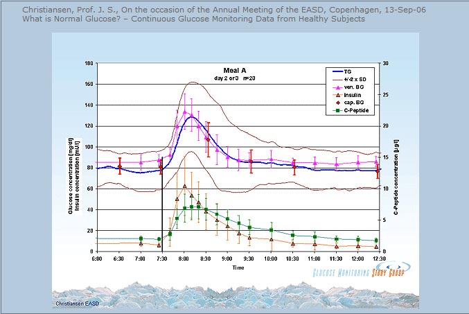
The issue of blood sugar levels and glycosylation appears to be one of level and duration of exposure. If we plot blood sugar over time and draw a graph, the area under the curve of the graph represents total exposure to  levels of blood sugar. If we draw a straight horizontal line under this curve that represents a toxic threshold (levels that result in glycosylation that exceed our ability to eliminate AGEs)  then the area of toxicity is equal to the area above the threshold line and below the curve of blood sugar. In theory then we should live a lifestyle (nutrition, sleep, exercise, stress reduction) that results in keeping our blood sugars as close as possible to the threshold of toxicity. The Whitehall study suggests that line would be drawn at 86 mg/dl. this discussion provides a conceptual framework. There is no proof of this argument, just data that support the concept that as blood sugars stay elevated above a certain level, this elevation increases the risk of disease. When we examine this argument in the light of evolutionary medicine/health it makes sense. Before the onset of agriculture we did not consume added sweeteners, refined carbohydrates, refined “vegetable” oils (oils from seeds, grains and legumes), nor did we consume manufactured trans fats. So draw a horizontal line in the graph below at some level, make it 86 mg/dl, and look at the area between the blood sugar level and that horizontal line. That is the theoretical toxicity zone.

blood sugar curve

The association between “normal blood sugar levels” and risk of heart attack and stroke have been observed for a long time but this association has received much less attention than the concern over consumption of fat and cholesterol in the diet. In previous posts I have pointed out the evidence that contradicts the notion that  consumption of saturated fat and cholesterol is a problem. Instead, there is growing evidence that easily oxidized polyunsaturated fat (vegetable oil) contributes to atherosclerosis , cardiovascular disease and chronic inflammation. Likewise, there is growing evidence that consumption of sweetened foods and beverages, as well as refined flour foods (which increase blood sugars much more than whole foods) are wreaking havoc in many ways.

So if there is a link between dietary sugar/refined carbohydrate consumption, blood sugar levels and disease, mediated by inflammation and glycosylation, what can we do about it? If there is a link between excessive consumption of pro-inflammatory and easily oxidized refined vegetable oils (linoleic acid) what can we do about it?

  • Avoid sweetened food and beverages
  • Drink only water and modest amounts of coffee or tea.
  • Avoid flour foods and other forms of refined carbohydrate which result in blood sugar surges and over time stress the pancreas
  • Eat only whole foods
  • Save your carbs for dinner
  • Walk for 15 minutes after every meal or 30-45 minutes per day
  • Engage in resistance training (weight lifting, resistance bands) for 20-30 minutes twice per week.
  • Get 8-9 hours of sleep each night
  • And if you really want to get serious about nutritional changes,  eat only the foods we have evolved to eat. Eat like a hunter-gatherer. Eat only pastured meat, free range poultry and free range eggs, fresh  wild fish and seafood, fresh  vegetables, fresh fruits and nuts.  Avoid grains, legumes, dairy. Avoid refined vegetable oils. Do not eat any food with “partially hydrogenated oil” or “hydrogenated oil” of any kind.

Resistance training twice per week for just 20-30 minutes will increase muscle mass and insulin sensitivity, lower blood sugars, preserve bone density, and provide many health benefits.

Eating most  carbs at dinner improved weight loss, lowered hunger, reduced abdominal circumference and enhanced body fat mass reductions in a calorie restricted weight loss study of obese adults.

Greater weight loss and hormonal cha… [Obesity (Silver Spring). 2011] – PubMed – NCBI

Sleep deprivation impairs insulin sensitivity, increases the risk of diabetes, hypertension, cardiovascular disease, depression, accidents and cancer, impairs immune function and wound healing, and impairs weight loss on a calorie restricted diet.

Meta-Analysis of Short Sleep Duration and Obesity in Children and Adults

Sleep duration and body mass index in twins: a gene-en… [Sleep. 2012] – PubMed – NCBI

Impact of insufficient sleep on total daily energy expenditure, food intake, and weight gain.

Neurobiological consequences of sleep deprivation.

Sleep and type 2 diabetes mellitus- clinical implications.

The influence of shift work on cognitive functions and oxidative stress.

Sleep disorders and depression: brief review of the literature, case report, and nonpharmacologic interventions for depression.

The impact of sleep deprivation on food desire in the human brain.

Walking 15 minutes after every meal in adults 60 years and older significantly improved 24 hour blood glucose control relative to control subjects who did not walk and was significantly more effective than 45 minutes of sustained morning or afternoon walking in lowering 3 hour post-dinner glucose levels.

Three 15-min bouts of moderate postmeal walking significantly improves 24-h glycemic control in older people at risk for impaired glucose tolerance.

Food, sleep, exercise and stress are the primary determinants of health.

While this post discussed two of three proposed mechanisms linking blood sugar levels to disease (modified LDL and AGEs) I did not discuss inflammation. The relationship between dietary sugar, refined carbohydrates and inflammation will be discussed in future posts.

In the meantime, stay tuned for “an egg a day keeps the doctor away”.

Peace,

Bob Hansen MD